
- Submit a Protocol
- Receive Our Alerts
- EN

- Protocols

- Articles and Issues
- For Authors
- About
- Become a Reviewer

Current Issue in 2024
Volume: 14, Issue: 21
Biochemistry
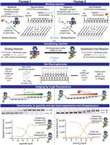
A Native PAGE Assay for the Biochemical Characterization of G Protein Coupling to GPCRs
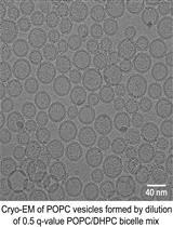
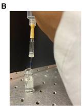
A Simple and Straightforward Approach for Generating Small, Stable, Homogeneous, Unilamellar 1-Palmitoyl 2-Oleoyl Phosphatidylcholine (POPC) Bilayer Vesicles
Biophysics
Single Molecular Resolution to Monitor DNA Replication Fork Dynamics upon Stress by DNA Fiber Assay
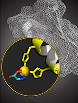
Copper Based Site-directed Spin Labeling of Proteins for Use in Pulsed and Continuous Wave EPR Spectroscopy

Label-free Quantification of Direct Protein-protein Interactions with Backscattering Interferometry

Cancer Biology
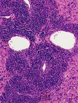
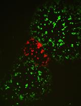
Reconstruction of Human AML Using Functionally and Immunophenotypically Defined Human Haematopoietic Stem and Progenitor Cells as Targeted Populations
Cell Biology
Preparation of Giant Escherichia coli spheroplasts for Electrophysiological Recordings
Developmental Biology
A Method to Induce Brown/Beige Adipocyte Differentiation from Murine Preadipocytes
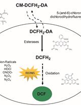
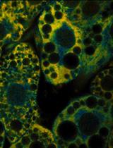
Measurement of Reactive Oxygen and Nitrogen Species in Living Cells Using the Probe 2',7'-Dichlorodihydrofluorescein
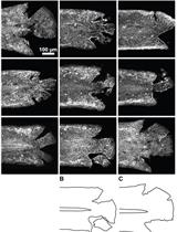
Wounding Zebrafish Larval Epidermis by Laceration
Microbiology
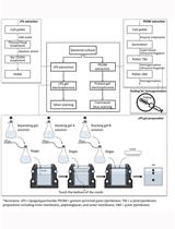
Extraction and Electrophoretic Analysis of Bacterial Lipopolysaccharides and Outer Membrane Proteins
Selection of Vaccinia Virus Recombinants Using CRISPR/Cas9
Neuroscience

Delivery of AAV for Expression of Fluorescent Biosensors in Juvenile Mouse Hippocampus
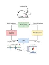
Assessment of Corticospinal Excitability in Awake Rodents Using EMG-Controlled Intracortical Stimulation

Plant Science
Non-invasive Imaging of Rice Roots in Non-compacted and Compacted Soil
Bacterial Infection and Hypersensitive Response Assays in Arabidopsis-Pseudomonas syringae Pathosystem
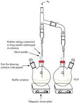
An in vitro Coupled Assay for PEPC with Control of Bicarbonate Concentration