
- Submit a Protocol
- Receive Our Alerts
- EN

- Protocols

- Articles and Issues
- For Authors
- About
- Become a Reviewer

Past Issue in 2014
Volume: 4, Issue: 14
Cancer Biology
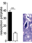
Bone Resorption Assay

Developmental Biology
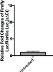
Wnt Reporter Activity Assay

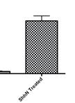
Hedgehog (Hh) Reporter Activity Assay
Fluorescence Microscopy for Cilia in Cultured Cells and Zebrafish Embryos

Immunology
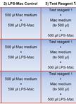
Macrophage Inflammatory Assay


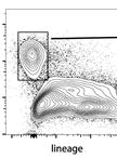
Isolation of ILC2 from Mouse Liver


Microbiology
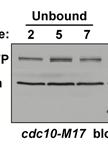
Chromatin Fractionation Assay in Fission Yeast
Dye Release Experiments with Dextran Loaded Vesicles

Development of a Novel Assay for Synthesis and Hydrolysis of Sedoheptulose 1,7-bisphosphate (SBP) in vitro by Combinations of Purified Fructose 1,6-bisphosphate aldolases (FBA) Proteins and Fructose 1,6-bisphosphatases (FBPase) Proteins from Bacillus methanolicus MGA3


Bacterial Fluorescent-dextran Diffusion Assay
Affinofile Assay for Identifying Macrophage-Tropic HIV-1
In vitro EBV Infection of Mononuclear Cells that Have Been Cryo-preserved

Plant Science
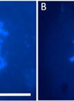
Rice Meiotic Chromosome Spread Preparation of Pollen Mother Cells
Stem Cell
Mesenchymal Stem Cell (MSC) Aggregate Formation in vivo
Coculture between hMADS and Mouse Adult CM


