
- Submit a Protocol
- Receive Our Alerts
- EN

- Protocols

- Articles and Issues
- For Authors
- About
- Become a Reviewer

Past Issue in 2014
Volume: 4, Issue: 17
Biochemistry
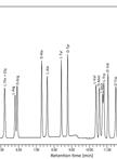
Analysis of L- and D-Amino Acids Using UPLC
Cell Biology
Dye-uptake Experiment through Connexin Hemichannels


Microbiology
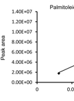
Metabolite and Fatty Acid Analysis of Yeast Cells and Culture Supernatants


Iodine Staining of Escherichia coli Expressing Genes Involved in the Synthesis of Bacterial Glycogen


Fitness Measurements of Evolved Esherichia coli


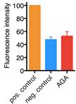
Evolution of Escherichia coli to Macrophage Cell Line
Purification of the GfsA-3x FLAG Protein Expressed in Aspergillus nidulans

Induction of Connexin-hemichannel Opening
Determination of D-galactofuranose Content of Galactomannoproteins in Aspergillus nidulans
Plant Science
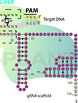
Targeted Gene Mutation in Rice Using a CRISPR-Cas9 System


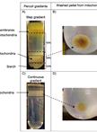
Isolation of Mitochondria from Potato Tubers

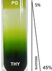
Gradient Flotation Centrifugation of Chloroplast Membranes

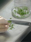
Axenic Culture of Sclerotinia sclerotiorum and Preparation of Sclerotinia Culture Filtrate Elicitor 1 (SCFE1)-containing Fractions, Triggering Immune Responses in Arabidopsis thaliana

Centrifuge Microscopy to Analyze the Sedimentary Movements of Amyloplasts