
- Submit a Protocol
- Receive Our Alerts
- EN

- Protocols

- Articles and Issues
- For Authors
- About
- Become a Reviewer

Past Issue in 2014
Volume: 4, Issue: 18
Biochemistry
Dimethylmethylene Blue Assay (DMMB)
Protein-lipid Interaction Analysis by Surface Plasmon Resonance (SPR)


Immunology
Isolation and FACS Analysis on Mononuclear Cells from CNS Tissue
Generation of Aβ-specific T cell lines and in vivo Transfer
Microbiology
Gentamicin Protection Assay to Determine Bacterial Survival within Macrophages
In vitro Transcription (IVT) and tRNA Binding Assay


Activity Assays for Bacteriophage Endolysin PlyPy


Detection of Hog1 Phosphorylation in Candida albicans in Response to an Antifungal Protein
Protocol for Biotin Bioassay-based Cross Feeding

Neuroscience
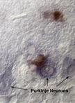
Combined in situ Hybridization/Immunohistochemistry (ISH/IH) on Free-floating Vibratome Tissue Sections

Plant Science
Perls Staining for Histochemical Detection of Iron in Plant Samples


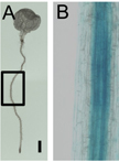
Extraction of Chloroplast Proteins from Transiently Transformed Nicotiana benthamiana Leaves
Thin Sections of Technovit 7100 Resin of Rice Endosperm and Staining
