
- Submit a Protocol
- Receive Our Alerts
- EN

- Protocols

- Articles and Issues
- For Authors
- About
- Become a Reviewer

Past Issue in 2014
Volume: 4, Issue: 20
Immunology
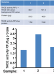
Measurement of TACE Activity in Extracts from Cultured Cells


Microbiology
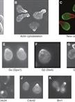
Synchronization of Saccharomyces cerevisiae Cells in G1 Phase of the Cell Cycle


Extraction and Purification of Mycobacterial Mycolic Acids
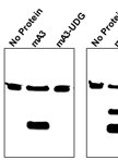
In vitro Assay for Cytidine Deaminase Activity of APOBEC3 Protein


Measuring UV-induced Mutagenesis at the CAN1 Locus in Saccharomyces cerevisiae
Design of a Transcription-based Secretion Activity Reporter (TSAR) for the Type III Secretion Apparatus of Shigella flexneri and Uses Thereof


Detection of the Secreted and Cytoplasmic Fractions of IpaB, IpaC and IpaD by Lysozyme Permeabilization
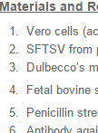
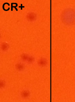
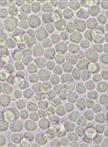
Severe Fever with Thrombocytopenia Syndrome Virus Infection of Cell Cultures


Molecular Biology
Expression and Purification of the Eukaryotic MBP-MOS1 Transposase from Sf21 Insect Cells

Neuroscience
Olfactory Habituation in Fasted Mice


Plant Science
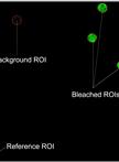
Fluorescence Recovery after Photobleaching (FRAP) Assay to Measure the Dynamics of Fluorescence Tagged Proteins in Endoplasmic Reticulum Membranes of Plant Cells


Plastic Embedding of Arabidopsis Stem Sections

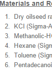
Seed Storage Reserve Analysis

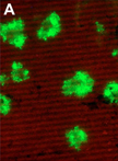
A Protocol to Measure the Extent of Cell-to-cell Movement of RNA Viruses in Planta