
- Submit a Protocol
- Receive Our Alerts
- EN

- Protocols

- Articles and Issues
- For Authors
- About
- Become a Reviewer

Past Issue in 2014
Volume: 4, Issue: 22
Cancer Biology
Purification of Tumor-Associated Macrophages (TAM) and Tumor-Associated Dendritic Cells (TADC)
Induction of Colitis and Colitis-associated Colorectal Cancer (CAC)
Cell Biology
Analysis of Intestinal Permeability in Mice
Separation of the Inner and Outer Mitochondrial Membrane in HeLa Cells
Assay of Ornithine Decarboxylase and Spermidine/Spermine N1-acetyltransferase Activities


Preparation of Golgi Membranes from Rat Liver
Microbiology
Infectious Focus Assays and Multiplicity of Infection (MOI) Calculations for Alpha-herpesviruses

Semi-denaturing Detergent Agarose Gel Electrophoresis (SDD-AGE)


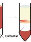
Isolation and Characterisation of Dendritic Cells from Peripheral Blood
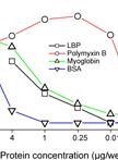
Competitive ELISA for Protein-lipopolysaccharide (LPS) Binding

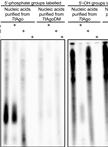
Purification and Sequencing of DNA Guides from Prokaryotic Argonaute


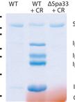
Secretion Assay in Shigella flexneri
Plant Science
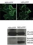
In vitro Detection of S-acylation on Recombinant Proteins via the Biotin-Switch Technique

Stem Cell
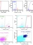
Isolation of FAP Cells from Mouse Dystrophic Skeletal Muscle Using Fluorescence Activated Cell Sorting

