Past Issue in 2014

Volume: 4, Issue: 24

left

Dec

20

Dec

5

Nov

20

Nov

5

Oct

20

Oct

5

Sep

20

Sep

5

Aug

20

Aug

5

Jul

20

Jul

5

Jun

20

Jun

5

May

20

May

5

Apr

20

Apr

5

Mar

20

Mar

5

Feb

20

Feb

5

Jan

20

Jan

5

right

Biochemistry

RNA Chromatin Immunoprecipitation (RNA-ChIP) in Caenorhabditis elegans

RNA Chromatin Immunoprecipitation (RNA-ChIP) in Caenorhabditis elegans

Germano Cecere Germano Cecere
Alla Grishok Alla Grishok
14132 Views
Dec 20, 2014
The RNA chromatin immunoprecipitation assay (RNA-ChIP) allows detection and quantification of RNA–protein interactions using in vivo cross-linking with formaldehyde followed by immunoprecipitation of the RNA–protein complexes. Here we describe the RNA–ChIP protocol that we have adapted for Caenorhabditis elegans (C. elegans) to detect interaction between the nuclear Argonaute CSR-1 (chromosome segregation and RNAi deficient) protein and its target nascent RNAs. We have used a transgenic strain expressing a recombinant long isoform of CSR-1 protein fused with N-terminal 3x FLAG epitope.

Microbiology

Fluorescence-linked Antigen Quantification (FLAQ) Assay for Fast Quantification of HIV-1 p24Gag

Fluorescence-linked Antigen Quantification (FLAQ) Assay for Fast Quantification of HIV-1 p24Gag

MG Marianne Gesner
MM Mekhala Maiti
RG Robert Grant
Marielle  Cavrois Marielle Cavrois
10699 Views
Dec 20, 2014
The fluorescence-linked antigen quantification (FLAQ) assay allows a fast quantification of HIV-1 p24Gag antigen. Viral supernatant are lysed and incubated with polystyrene microspheres coated with polyclonal antibodies against HIV-1 p24Gag and detector antibodies conjugated to fluorochromes (Figure 1). After washes, the fluorescence of microspheres is measured by flow cytometry and reflects the abundance of the antigen in the lysate. The speed, simplicity, and wide dynamic range of the FLAQ assay are optimum for many applications performed in HIV-1 research laboratories.
Hepatitis C virus Cell-to-cell Spread Assay

Hepatitis C virus Cell-to-cell Spread Assay

Naina Barretto Naina Barretto
Susan L. Uprichard Susan L. Uprichard
10534 Views
Dec 20, 2014
Hepatitis C virus (HCV) can infect naïve cells via entry of “cell-free” extracellular virus or direct “cell-to-cell” transmission. Here, we describe an assay for detecting HCV cell-to-cell transmission, using a non-growing cell culture system that avoids confounding effects of cell growth. The assay consists of infecting a small number of cells in a confluent monolayer and then blocking subsequent cell-free extracellular virions with a neutralizing antibody such that only cell-to-cell transmission may occur. Under these conditions, incubation at 37 °C results in the formation of infected cell foci. The extent of cell-to-cell spread can then be determined by counting the number of cells in each focus. The assay may be modified to assess the effects of inhibitors and/or specific cellular genes on cell-to-cell spread of HCV.

Plant Science

Virus-induced Gene Silencing (VIGS) in Phalaenopsis Orchids

Virus-induced Gene Silencing (VIGS) in Phalaenopsis Orchids

Ming-Hsien Hsieh Ming-Hsien Hsieh
ZP Zhao-Jun Pan
Hsin-Hung Yeh Hsin-Hung Yeh
Hong-Hwa Chen Hong-Hwa Chen
12447 Views
Dec 20, 2014
This is a protocol to produce stable silencing efficacy and efficiency for VIGS using CymMV as a silencing vector for floral functional genomics in Phalaenopsis orchids. This protocol is established based on a method created by Lu et al. (2007), and then modified by Hsieh et al. (2013a; 2013b), Lu et al. (2012) successfully engineered a cloning vector (pCymMV-Gateway) in that the target gene fragment is simple to insert and can be manipulated with high efficiency. The silencing vector is inoculated into plants by Agro-inoculation by using Agrobacterium tumefaciens (A. tumefaciens) strain EHA105. Agro-infiltration of leaves for use in VIGS study of orchid flowers is a time saver and produces less damage to flower buds.
Localization and Topology of Thylakoid Membrane Proteins in Land Plants

Localization and Topology of Thylakoid Membrane Proteins in Land Plants

ST Salar Torabi
MP Magdalena Plöchinger
JM Jörg Meurer
12152 Views
Dec 20, 2014
Thylakoids are a formation of flattened membrane vesicles and protein complexes found in cyanobacteria, algae and plants. In the chloroplasts of land plants the thylakoid membrane systems form a network of densely packed stacks called grana lamellae, which are connected by unstacked stroma lamellae. Photosystem II is mainly localized in the appressed grana region, while photosystem I and the ATP synthase complexes are enriched in the stroma lamellae. The cytochrome b6/f complex is distributed laterally throughout both stacked and unstacked membrane regions. The photosynthetic complexes consist of integral and peripheral proteins. The first part of this protocol (A) shows how to fractionate thylakoids into grana and stroma lamellae. The second part of this protocol (B) shows how to distinguish between strong hydrophobic integral membrane associations and weak electrostatic membrane and/or membrane complex associations. As it is necessary to specifically detect the protein of interest in the fractions, a specific antibody raised against the protein of interest or a complemented null mutant of a structural component expressing a tagged fusion protein would be of great advantage. The last part of this protocol (C) shows, how to investigate the topology of integral and peripheral proteins. This method requires a specific antibody for the protein of interest. For integral membrane proteins peptide-specific antibodies or epitope-tagged versions are required. The protocol is suitable for the investigation of low molecular weight proteins (LMW) below 5 kDa (Torabi et al., 2014).
Imaging and Measurement of Nanomechanical Properties within Primary Xylem Cell Walls of Broadleaves

Imaging and Measurement of Nanomechanical Properties within Primary Xylem Cell Walls of Broadleaves

Jaroslav Ďurkovič Jaroslav Ďurkovič
MK Monika Kardošová
RL Rastislav Lagaňa
11936 Views
Dec 20, 2014
A technique of atomic force microscopy (AFM) called PeakForce quantitative nanomechanical mapping (PeakForce QNM) is an efficient tool for the quantitative mechanobiological imaging of fibrillar aggregate, human epidermal cell and woody plant cell wall topography (Sweers et al., 2011; Heu et al., 2012; Ďurkovič et al., 2012; Ďurkovič et al., 2013). Here, we describe a detailed protocol for the measurement of nanomechanical properties of primary xylem cell walls in woody plants, for the determination of reduced Young’s modulus of elasticity (MOE), adhesion, deformation, and energy dissipation (Figure 1). This new technique provides direct control of the maximum loading force and the deformation depth in cell wall samples keeping indentations small, while at the same time eliminating damaging lateral forces in order to preserve both the AFM tip and plant sample. High-resolution and non-destructive imaging shed new quantitative mechanistic insights into the structural biology of woody plant cell walls. This procedure can also be adapted for other biological samples with a varying range of stiffness.
Polysomal-mRNA Extraction from Arabidopsis by Sucrose-gradient Separation

Polysomal-mRNA Extraction from Arabidopsis by Sucrose-gradient Separation

SL Shih-Yun Lin
Guang-Yuh Jauh Guang-Yuh Jauh
11539 Views
Dec 20, 2014
mRNAs surrounded by polysomes are ready for translation into proteins (Warner et al., 1963); these mRNAs are defined as polysomal-mRNAs (Mustroph et al., 2009). The process is affected by various growth conditions or surrounding situations. Microarray analysis is a powerful tool for detecting genome-wide gene expression. Therefore, using polysomal-mRNAs for microarray analysis can reflect the gene translation information (the translatome) under different developmental stages or environmental conditions from eukaryotes. Polysomal-mRNAs can be collected from the polysomal fraction by sucrose-gradient separation for further quantitative PCR or microarray assay. We modified a protocol (Mustroph et al., 2009) for collecting polysomal-mRNAs via sucrose-gradient separation to eliminate monosomal-mRNA contamination from pLAT52:HF:RPL18 Arabidopsis. This transgenic Arabidopsis uses a pollen-specific promoter (ProLAT52) to generate epitope-tagged polysomal-RNA complexes that could be purified with a specific antigen (Lin et al., 2014). The polysomal-mRNAs we obtained via sucrose-gradient separation and antibody purification underwent in vivo translation in pollen tubes grown from self-pollinated gynoecia of Arabidopsis thaliana.
Cyclohexane Diamine Tetraacetic Acid (CDTA) Extraction of Plant Cell Wall Pectin

Cyclohexane Diamine Tetraacetic Acid (CDTA) Extraction of Plant Cell Wall Pectin

GB Gerit Bethke
JG Jane Glazebrook
11518 Views
Dec 20, 2014
The goal of this procedure is to extract pectin from plant cell walls. Pectins are galacturonic acid containing polymeric sugars that are important components of plant cell walls. Various procedures aimed at studying plant cell wall components require the extraction of pectin. Pectin is synthesized in the Golgi apparatus in a highly esterified fashion and is de-esterified in the cell wall (Mohnen, 2008). Pectin is generally water soluble. De-esterified pectin can form so-called “egg-box structures” in the presence of Ca2+ ions (Mohnen, 2008; Harholt et al., 2010). Pectin in these “egg-box structures” is cross-linked and less soluble. Cyclohexane diamine tetraacetic acid (CDTA) chelates Ca2+ ions and hence allows extraction of Ca2+ cross-linked pectin from cell walls.
Agrobacterium-mediated Transformation of Mature Ginseng Embryos

Agrobacterium-mediated Transformation of Mature Ginseng Embryos

OL Ok Ran Lee
JH Jin Haeng Han
YK Yoon-Ri Kim
11229 Views
Dec 20, 2014
Ginseng refers to species within the genus Panax and is a slow-growing perennial herb from the Araliaceae family. The most widely used Panax species is Panax ginseng Meyer (Korean ginseng). Panax japonicus (Japanese ginseng), Panax notoginseng (Chinese ginseng), and Panax quinquefolium (American ginseng). Due to the various pharmaceutical importance of ginsenosides, ginseng plant has been cultivated for its highly valued root over 2,000 years as a medicinal plant in East Asian countries particularly in China, Korea, and Japan and North America. Korean ginseng (Panax ginseng C. A. Meyer) consists of nine cultivars from three Jakyung, Chungkyung, and Hwangsook lines. Cultivar “Yunpoong” has characteristics to have more axillary shoots and lateral roots compared to other cultivars. Thus “Yunpoong”: Ginseng seeds are relatively more feasible for regeneration of adventitious roots during gene transformation. Here, we describe how to prepare and treat ginseng seeds after harvest till the ginseng immature embryos are ready for the germination, and to be used in Agrobacterium tumefaciens-mediated gene transformation.
Trichome Isolation and Integrity Test from Brassica villosa and Other Species

Trichome Isolation and Integrity Test from Brassica villosa and Other Species

NN Naghabushana Nayidu
PB Peta Bonham-Smith
MG Margaret Y. Gruber
9404 Views
Dec 20, 2014
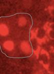
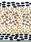
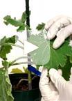
The outward growths from one or more epidermal cells are well known as trichomes (plant hair cells) (Levin, 1973; Mathur, 2006). Preparation of pure intact non-glandular trichomes from trichome-rich Brassica villosa depended on rendering the trichomes sufficiently stiff to dislodge them from the leaves in an undamaged state, which can be further used for structural, transcriptome, genome and biochemical or chemical analysis. Dislodging the flexible trichomes from Brassica villosa (B. villosa) with a paintbrush [as in Zhang and Oppenheimer (2004)] proved too gentle, and scraping trichomes off the leaves with a razor blade [as in Zhang and Oppenheimer (2004)] resulted in trichome cell disruption. Aziz et al. (2005) reported isolating alfalfa glandular trichomes by shearing in liquid nitrogen (N2). In the present study, a similar method was used. Non-glandular leaf trichomes were isolated by treating tissue with liquid N2 to stiffen the flexible trichomes, followed by 1 min of shearing force with a common vortex mixer to dislodge the trichomes from their leaf bed (Nayidu et al., 2014). Up-to ~20 % of the trichomes were removed from the leaf surface and the majority were unbroken (confirmed by no staining by trypan blue; Figure 1). Trichomes were then purified by straining a tissue/trichome/water mixture through a sieve. However, leaf tissues also became very brittle after dipping in liquid N2 and broke up into very small pieces if the leaf tissue was agitated for a longer time period. Hence, N2-treated leaf samples could not be re-used to recover remaining attached trichomes. Contaminating leaf pieces from a 1 min shear were larger and easily to separate manually from the detached trichomes within the sieve, rendering a purified intact trichome preparation. The method was also successful at purifying trichomes from soybean (Glycine max) and tomato (Solanum lycopersicum).