
- Submit a Protocol
- Receive Our Alerts
- EN

- Protocols

- Articles and Issues
- For Authors
- About
- Become a Reviewer

Past Issue in 2017
Volume: 7, Issue: 6
Biochemistry
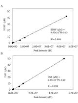
Measurement of RNA-induced PKR Activation in vitro

Cancer Biology

Polysome Analysis


RNA-protein UV-crosslinking Assay
3D Stroma Invasion Assay
Developmental Biology
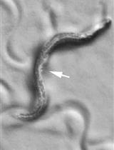
Measuring Caenorhabditis elegans Sleep during the Transition to Adulthood Using a Microfluidics-based System
Immunology
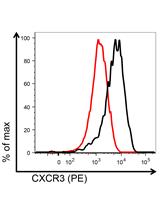
Mouse CD8+ T Cell Migration in vitro and CXCR3 Internalization Assays


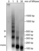
Differential Salt Fractionation of Nuclei to Analyze Chromatin-associated Proteins from Cultured Mammalian Cells
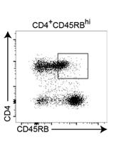
Mouse Model of Reversible Intestinal Inflammation
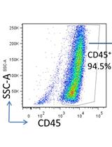
Adoptive Transfer of Lung Antigen Presenting Cells
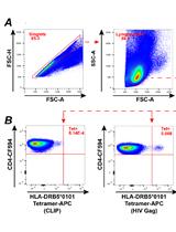
MHC Class II Tetramer Labeling of Human Primary CD4+ T Cells from HIV Infected Patients
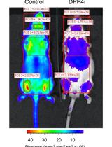
Measurement of Dipeptidylpeptidase Activity in vitro and in vivo

Microbiology
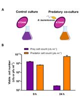
Determination of the Predatory Capability of Bdellovibrio bacteriovorus HD100
Wheat Root-dip Inoculation with Fusarium graminearum and Assessment of Root Rot Disease Severity


Extraction, Purification and Quantification of Diffusible Signal Factor Family Quorum-sensing Signal Molecules in Xanthomonas oryzae pv. oryzae


In vitro Assay to Assess Efficacy of Potential Antiviral Compounds against Enterovirus D68


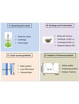
Isolation of Ribosomal Particles from the Unicellular Cyanobacterium Synechocystis sp. PCC 6803
Assays for the Detection of Rubber Oxygenase Activities

Determination of Adeno-associated Virus Rep DNA Binding Using Fluorescence Anisotropy

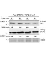
Chase Assay of Protein Stability in Haloferax volcanii

Molecular Biology
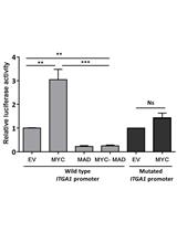
In silico Analysis and Site-directed Mutagenesis of Promoters


Plant Science
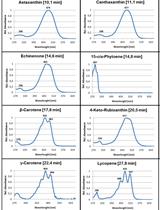
Extraction and Analysis of Carotenoids from Escherichia coli in Color Complementation Assays


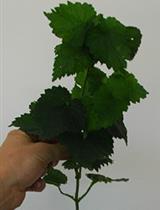
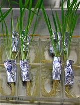
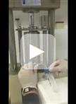
Xylem Sap Extraction Method from Hop Plants


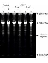
Ribosomal RNA N-glycosylase Activity Assay of Ribosome-inactivating Proteins


Rubisco Extraction and Purification from Diatoms

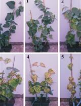
Pathogenicity Assay of Verticillium nonalfalfae on Hop Plants