
- Submit a Protocol
- Receive Our Alerts
- EN

- Protocols

- Articles and Issues
- For Authors
- About
- Become a Reviewer

Past Issue in 2017
Volume: 7, Issue: 8

Biochemistry
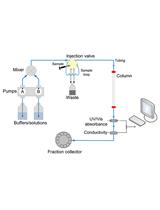
Determination of Hydrodynamic Radius of Proteins by Size Exclusion Chromatography
Expression and Purification of Mini G Proteins from Escherichia coli


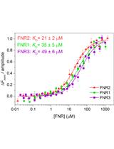
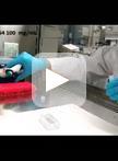
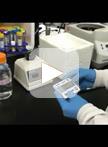
Measurement of FNR-NrdI Interaction by Microscale Thermophoresis (MST)
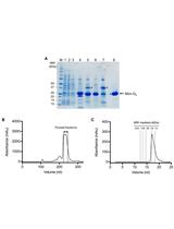
Expression, Purification and Crystallisation of the Adenosine A2A Receptor Bound to an Engineered Mini G Protein
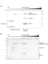
Affinity Purification of the RNA Degradation Complex, the Exosome, from HEK-293 Cells

Cancer Biology
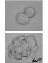
Melanoma Stem Cell Sphere Formation Assay

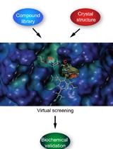
Virtual Screening of Transmembrane Serine Protease Inhibitors
Cell Biology
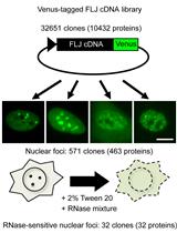
RNase Sensitivity Screening for Nuclear Bodies with RNA Scaffolds in Mammalian Cells
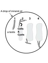
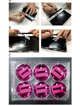
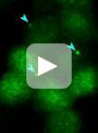
Time-lapse Observation of Chromosomes, Cytoskeletons and Cell Organelles during Male Meiotic Divisions in Drosophila
Reversible Cryo-arrests of Living Cells to Pause Molecular Movements for High-resolution Imaging


Developmental Biology
Physical Removal of the Midbody Remnant from Polarised Epithelial Cells Using Take-Up by Suction Pressure (TUSP)

Immunology
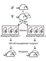
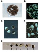
Lentiviral Barcode Labeling and Transplantation of Fetal Liver Hematopoietic Stem and Progenitor Cells
Microbiology

CRISPR/Cas9 Editing of the Bacillus subtilis Genome
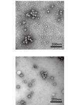
Isolation of the Dot/Icm Type IV Secretion System Core Complex from Legionella pneumophila for Negative Stain Electron Microscopy Studies

Molecular Biology
Expression and Analysis of Flow-regulated Ion Channels in Xenopus Oocytes
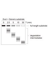
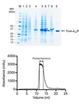
RNA Degradation Assay Using RNA Exosome Complexes, Affinity-purified from HEK-293 Cells
Neuroscience
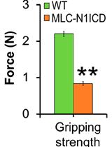
Evaluation of Muscle Performance in Mice by Treadmill Exhaustion Test and Whole-limb Grip Strength Assay
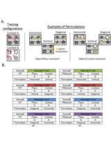
The Object Context-place-location Paradigm for Testing Spatial Memory in Mice


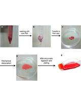
Preparation of Primary Astrocyte Culture Derived from Human Glioblastoma Multiforme Specimen
Plant Science
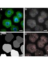
Single Molecule RNA FISH in Arabidopsis Root Cells

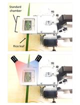
Measurement of Stomatal Conductance in Rice
Growth Assay for the Stem Parasitic Plants of the Genus Cuscuta

Automated Tracking of Root for Confocal Time-lapse Imaging of Cellular Processes


Rapid Isolation of Total Protein from Arabidopsis Pollen

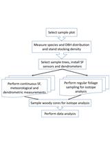
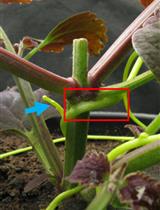
Forest GPP Calculation Using Sap Flow and Water Use Efficiency Measurements
Stem Cell
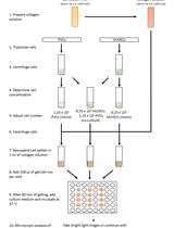
Mimicking Angiogenesis in vitro: Three-dimensional Co-culture of Vascular Endothelial Cells and Perivascular Cells in Collagen Type I Gels