
- Submit a Protocol
- Receive Our Alerts
- EN

- Protocols

- Articles and Issues
- For Authors
- About
- Become a Reviewer

Past Issue in 2017
Volume: 7, Issue: 13
Biochemistry
Active Cdk5 Immunoprecipitation and Kinase Assay
Formaldehyde Fixation of Extracellular Matrix Protein Layers for Enhanced Primary Cell Growth


Cancer Biology
Tumorigenicity Assay in Nude Mice

A Novel Mouse Skin Graft Model of Vascular Tumors Driven by Akt1

Generation of a Cellular Reporter for Functional BRD4 Inhibition

Cell Biology
Endpoint or Kinetic Measurement of Hydrogen Sulfide Production Capacity in Tissue Extracts
Photothrombotic Induction of Capillary Ischemia in the Mouse Cortex during in vivo Two-Photon Imaging
Developmental Biology

Oxidative Stress Assays (arsenite and tBHP) in Caenorhabditis elegans

Ex vivo Ooplasmic Extract from Developing Drosophila Oocytes for Quantitative TIRF Microscopy Analysis


Immunology
In vitro Demonstration and Quantification of Neutrophil Extracellular Trap Formation

EAE Induction by Passive Transfer of MOG-specific CD4+ T Cells
Qualitative and Quantitative Assay for Detection of Circulating Autoantibodies against Human Aortic Antigen
Vaginal HSV-2 Infection and Tissue Analysis
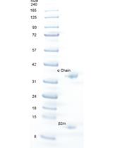
Thermal Stability of Heterotrimeric pMHC Proteins as Determined by Circular Dichroism Spectroscopy

Microbiology
Assaying Glycogen and Trehalose in Yeast
Bacterial Survival in Dictyostelium

Determination of NO and CSF Levels Produced by Bacillus subtilis


Molecular Biology
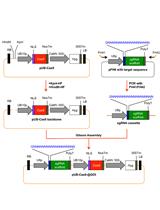
Multiplex Gene Editing via CRISPR/Cas9 System in Sheep
Neuroscience
Spinal Cord Preparation from Adult Red-eared Turtles for Electrophysiological Recordings during Motor Activity


Behavioral and Functional Assays for Investigating Mechanisms of Noxious Cold Detection and Multimodal Sensory Processing in Drosophila Larvae


Live Imaging of Myogenesis in Indirect Flight Muscles in Drosophila
Delayed-matching-to-place Task in a Dry Maze to Measure Spatial Working Memory in Mice
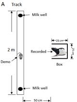
Social Observation Task in a Linear Maze for Rats
Plant Science
Generation of Targeted Knockout Mutants in Arabidopsis thaliana Using CRISPR/Cas9

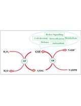
Determination of Reduced and Total Glutathione Content in Extremophilic Microalga Galdieria phlegrea


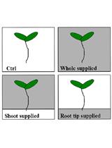
Determination of the Effects of Local and Systemic Iron Excess on Lateral Root Initiation in Arabidopsis thaliana

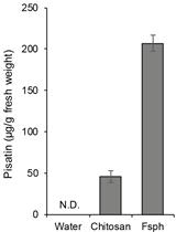
A Simple and Rapid Assay for Measuring Phytoalexin Pisatin, an Indicator of Plant Defense Response in Pea (Pisum sativum L.)
Stem Cell
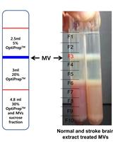
Microvesicle Isolation from Rat Brain Extract Treated Human Mesenchymal Stem Cells