
- Submit a Protocol
- Receive Our Alerts
- EN

- Protocols

- Articles and Issues
- For Authors
- About
- Become a Reviewer

Past Issue in 2017
Volume: 7, Issue: 14
Biochemistry
Cell Type-specific Metabolic Labeling of Proteins with Azidonorleucine in Drosophila

Mass Spectrometry-based in vitro Assay to Identify Drugs that Influence Cystine Solubility


In vitro AMPylation Assays Using Purified, Recombinant Proteins
![[2-3H]Mannose-labeling and Analysis of N-linked Oligosaccharides](https://en-cdn.bio-protocol.org/imageup/arcimg/20170718074630401.jpg?t=1758530367)
[2-3H]Mannose-labeling and Analysis of N-linked Oligosaccharides

Cancer Biology
Measurement of the Intracellular Calcium Concentration with Fura-2 AM Using a Fluorescence Plate Reader
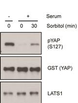
Non-radioactive LATS in vitro Kinase Assay
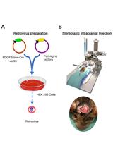
Glioma Induction by Intracerebral Retrovirus Injection


Immunology
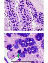
Analyzing the Properties of Murine Intestinal Mucins by Electrophoresis and Histology


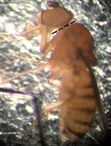
Preparation of Mosquito Salivary Gland Extract and Intradermal Inoculation of Mice

Microbiology
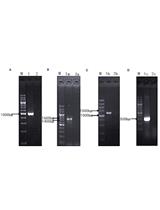
Using CRISPR/Cas9 for Large Fragment Deletions in Saccharomyces cerevisiae
Quantification of Chlorophyll as a Proxy for Biofilm Formation in the Cyanobacterium Synechococcus elongatus
Bioelectrospray Methodology for Dissection of the Host-pathogen Interaction in Human Tuberculosis
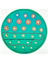
Xanthoferrin Siderophore Estimation from the Cell-free Culture Supernatants of Different Xanthomonas Strains by HPLC


Plasmodium Sporozoite Motility on Flat Substrates
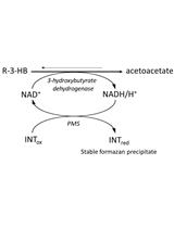
Quantitative Determination of Poly-β-hydroxybutyrate in Synechocystis sp. PCC 6803
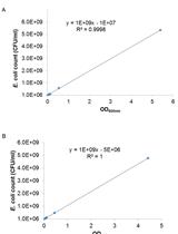
Determination of Survival of Wildtype and Mutant Escherichia coli in Soil
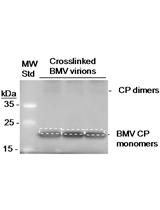
Mapping RNA Sequences that Contact Viral Capsid Proteins in Virions

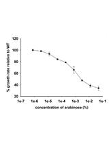
Gene Dosage Experiments in Enterobacteriaceae Using Arabinose-regulated Promoters
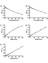
The Sulfur Oxygenase Reductase Activity Assay: Catalyzing a Reaction with Elemental Sulfur as Substrate at High Temperatures
Neuroscience

Aldicarb-induced Paralysis Assay to Determine Defects in Synaptic Transmission in Caenorhabditis elegans

Ciberial Muscle 9 (CM9) Electrophysiological Recordings in Adult Drosophila melanogaster

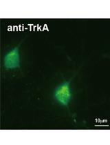
Isolation and Culturing of Rat Primary Embryonic Basal Forebrain Cholinergic Neurons (BFCNs)

Plant Science
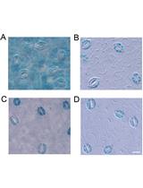
GUS Staining of Guard Cells to Identify Localised Guard Cell Gene Expression
Rice Lamina Joint Inclination Assay

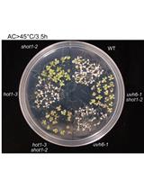
Assessing Plant Tolerance to Acute Heat Stress
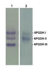
Separation of Plant 6-Phosphogluconate Dehydrogenase (6PGDH) Isoforms by Non-denaturing Gel Electrophoresis


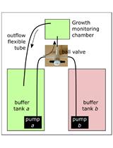
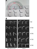
Non-invasive Protocol for Kinematic Monitoring of Root Growth under Infrared Light
Isolation of Fucus serratus Gametes and Cultivation of the Zygotes

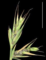
Isolation of Ustilago bromivora Strains from Infected Spikelets through Spore Recovery and Germination
Stem Cell

Implantation of Human Peripheral Corneal Spheres into Cadaveric Human Corneal Tissue