
- Submit a Protocol
- Receive Our Alerts
- EN

- Protocols

- Articles and Issues
- For Authors
- About
- Become a Reviewer

Past Issue in 2017
Volume: 7, Issue: 15
Biochemistry
Separation and Purification of Glycosaminoglycans (GAGs) from Caenorhabditis elegans

Purification of FLAG-tagged Secreted Proteins from Mammalian Cells
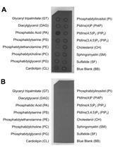
Membrane Lipid Screen to Identify Molecular Targets of Biomolecules
Isolation of Keratan Sulfate Disaccharide-branched Chondroitin Sulfate E from Mactra chinensis
Cancer Biology
RNA Interference Screening to Identify Proliferation Determinants in Breast Cancer Cells
Microbiology
CRISPR/Cas9 Gene Editing in the Marine Diatom Phaeodactylum tricornutum

Liposome Disruption Assay to Examine Lytic Properties of Biomolecules
Digestion of Peptidoglycan and Analysis of Soluble Fragments


A Protocol of Using White/Red Color Assay to Measure Amyloid-induced Oxidative Stress in Saccharomyces cerevisiae

Selection of Genetically Modified Bacteriophages Using the CRISPR-Cas System

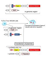
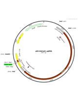
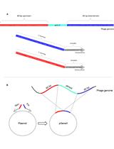
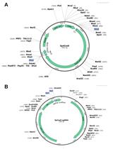
Advanced Design of Minimalistic Dumbbell-shaped Gene Expression Vectors


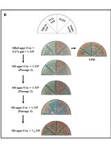
Observation of Pneumococcal Phase Variation in Colony Morphology


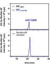
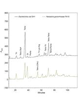
Analysis of N-acetylmuramic acid-6-phosphate (MurNAc-6P) Accumulation by HPLC-MS
Measurement of Energy-dependent Rhodamine 6G Efflux in Yeast Species
Molecular Biology
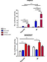
Improving CRISPR Gene Editing Efficiency by Proximal dCas9 Targeting
.jpg)
Neuroscience
Extraction of Soluble and Insoluble Protein Fractions from Mouse Brains and Spinal Cords

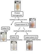
Preparation of Crude Synaptosomal Fractions from Mouse Brains and Spinal Cords
Plant Science
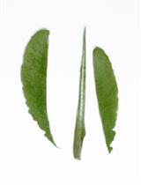
Isolation of Guard-cell Enriched Tissue for RNA Extraction


Isolation of Cytosol, Microsome, Free Polysomes (FPs) and Membrane-bound Polysomes (MBPs) from Arabidopsis Seedlings


Wheat Coleoptile Inoculation by Fusarium graminearum for Large-scale Phenotypic Analysis

Overrepresentation Analyses of Differentially Expressed Genes in the Smut Fungus Ustilago bromivora during Saprophytic and in planta Growth
Polyamine and Paraquat Transport Assays in Arabidopsis Seedling and Callus

Stem Cell
Differentiation of Human Induced Pluripotent Stem Cells (iPS Cells) and Embryonic Stem Cells (ES Cells) into Dendritic Cell (DC) Subsets

Mouse Müller Cell Isolation and Culture

Primary Culture System for Germ Cells from Caenorhabditis elegans Tumorous Germline Mutants