
- Submit a Protocol
- Receive Our Alerts
- EN

- Protocols

- Articles and Issues
- For Authors
- About
- Become a Reviewer

Past Issue in 2017
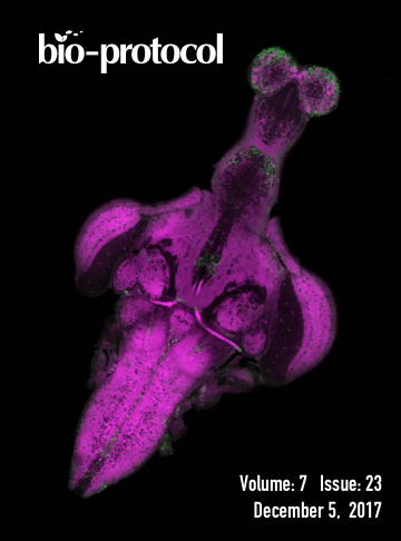
Volume: 7, Issue: 23
Cancer Biology
In vivo Leukemogenesis Model Using Retrovirus Transduction
Myeloid Progenitor Transformation Assay
Developmental Biology
Notch Ligand Binding Assay Using Flow Cytometry

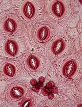
Mandibular Explant Assay for Investigating Extrinsic Stimuli on Bone and Cartilage Development


Immunology
Measuring the Endocytic Recycling of Amyloid Precursor Protein (APP) in Neuro2a Cells


Drosophila Model of Leishmania amazonensis Infection
Microbiology
Genome Editing in Diatoms Using CRISPR-Cas to Induce Precise Bi-allelic Deletions
Isolation, BODIPY Labeling and Uptake of Exosomes in Hepatic Stellate Cells

Lentiviral Knockdown of Transcription Factor STAT1 in Peromyscus leucopus to Assess Its Role in the Restriction of Tick-borne Flaviviruses
Brief Protocol for EDGE Bioinformatics: Analyzing Microbial and Metagenomic NGS Data
Determination of Mn Concentrations in Synechocystis sp. PCC6803 Using ICP-MS
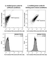
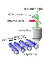
Quantification of Bacterial Polyhydroxybutyrate Content by Flow Cytometry
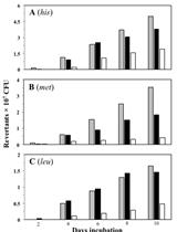
Stationary-phase Mutagenesis Soft-agar Overlay Assays in Bacillus subtilis
.jpg)

.jpg)
Molecular Biology
MicroScale Thermophoresis as a Tool to Study Protein-peptide Interactions in the Context of Large Eukaryotic Protein Complexes
Neuroscience
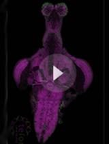
zPACT: Tissue Clearing and Immunohistochemistry on Juvenile Zebrafish Brain


Formalin Murine Model of Pain

Live Imaging of Axonal Transport in the Motor Neurons of Drosophila Larvae
 2.jpg)
A Novel Protocol to Quantitatively Measure the Endocytic Trafficking of Amyloid Precursor Protein (APP) in Polarized Primary Neurons with Sub-cellular Resolution
![Proboscis Extension Reflex in Apis mellifera [Honeybee] with Only One Antenna](https://en-cdn.bio-protocol.org/imageup/arcimg/20171129091737284.jpg?t=1758510586)
Proboscis Extension Reflex in Apis mellifera [Honeybee] with Only One Antenna

Plant Science
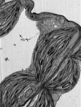
Using Light and Electron Microscopy to Estimate Structural Variation in Thylakoid Membranes
Automatic Leaf Epidermis Assessment Using Fourier Descriptors in Texture Images
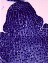
Investigating the Shape of the Shoot Apical Meristem in Bamboo Using a Superellipse Equation

Stem Cell
GC/MS-based Analysis of Volatile Metabolic Profile Along in vitro Differentiation of Human Induced Pluripotent Stem Cells


Systems Biology
Low-input Capture-C: A Chromosome Conformation Capture Assay to Analyze Chromatin Architecture in Small Numbers of Cells