
- Submit a Protocol
- Receive Our Alerts
- EN

- Protocols

- Articles and Issues
- For Authors
- About
- Become a Reviewer

Past Issue in 2018
Volume: 8, Issue: 5
Developmental Biology

Terminal Deoxynucleotidyl Transferase Mediated Production of Labeled Probes for Single-molecule FISH or RNA Capture


Immunology

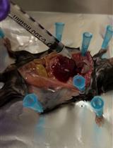
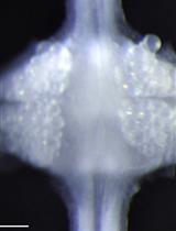
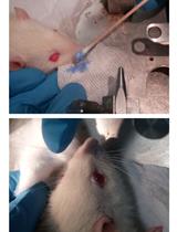
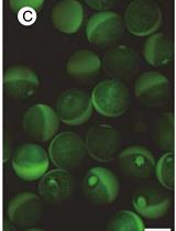
Murine Pancreatic Islets Transplantation under the Kidney Capsule


Mono Sodium Urate Crystal-induced Peritonitis for in vivo Assessment of Inflammasome Activation


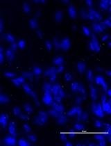
Quantification of Bacterial Attachment to Tissue Sections
Intravenous Labeling and Analysis of the Content of Thymic Perivascular Spaces
Microbiology
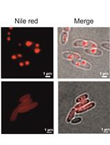
Determination of Polyhydroxybutyrate (PHB) Content in Ralstonia eutropha Using Gas Chromatography and Nile Red Staining
Visualization of RNA 3’ ends in Escherichia coli Using 3’ RACE Combined with Primer Extension
In vivo Analysis of Cyclic di-GMP Cyclase and Phosphodiesterase Activity in Escherichia coli Using a Vc2 Riboswitch-based Assay


Neuroscience
Barnes Maze Procedure for Spatial Learning and Memory in Mice
Construction and Cloning of Minigenes for in vivo Analysis of Potential Splice Mutations
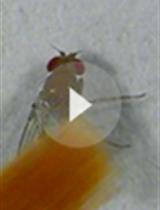
Flight and Climbing Assay for Assessing Motor Functions in Drosophila
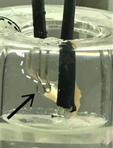
Mouse Phrenic Nerve Hemidiaphragm Assay (MPN)


Dual-sided Voltage-sensitive Dye Imaging of Leech Ganglia

A Fluorescent Dye Method Suitable for Visualization of One or More Rat Whiskers

Plant Science
Quantification of Plant Cell Death by Electrolyte Leakage Assay

Large Scale Field Inoculation and Scoring of Maize Southern Leaf Blight and Other Maize Foliar Fungal Diseases

Boron Uptake Assay in Xenopus laevis Oocytes

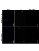
Histone Deubiquitination Assay in Nicotiana benthamiana


Systems Biology
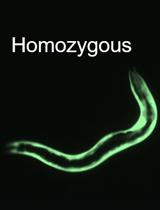
Synthetic Genetic Interaction (CRISPR-SGI) Profiling in Caenorhabditis elegans
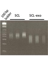
Coupling Exonuclease Digestion with Selective Chemical Labeling for Base-resolution Mapping of 5-Hydroxymethylcytosine in Genomic DNA
