
- Submit a Protocol
- Receive Our Alerts
- EN

- Protocols

- Articles and Issues
- For Authors
- About
- Become a Reviewer

Past Issue in 2018
Volume: 8, Issue: 8
Biochemistry
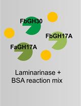
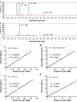
Laminarin Quantification in Microalgae with Enzymes from Marine Microbes

Cancer Biology
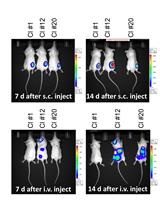
Generation of Luciferase-expressing Tumor Cell Lines
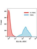
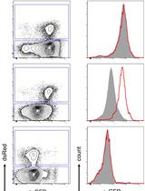
FACS-based Glucose Uptake Assay of Mouse Embryonic Fibroblasts and Breast Cancer Cells Using 2-NBDG Probe

Cell Biology
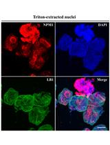
A Method for Extracting the Nuclear Scaffold from the Chromatin Network
Immunology
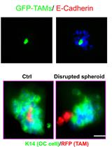
3D Co-culture System of Tumor-associated Macrophages and Ovarian Cancer Cells

Microbiology
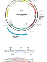
Method for CRISPR/Cas9 Mutagenesis in Candida albicans
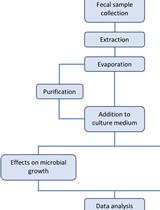
Extraction of Small Molecules from Fecal Samples and Testing of Their Activity on Microbial Physiology

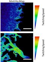
Quantification of Bacterial Twitching Motility in Dense Colonies Using Transmitted Light Microscopy and Computational Image Analysis


Metal-tagging Transmission Electron Microscopy for Localisation of Tombusvirus Replication Compartments in Yeast

Host-regulated Hepatitis B Virus Capsid Assembly in a Mammalian Cell-free System
Adhesion of Enteroaggregative E. coli Strains to HEK293 Cells
Determination of Intracellular Osmolytes in Cyanobacterial Cells
Molecular Biology
Generation of microRNA Sponge Library

Neuroscience
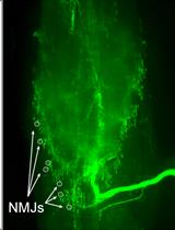
Electrophysiological Recordings of Evoked End-Plate Potential on Murine Neuro-muscular Synapse Preparations


Magnetic Resonance Imaging and Histopathological Visualization of Human Dural Lymphatic Vessels
Isolation and Maintenance of Murine Embryonic Striatal Neurons
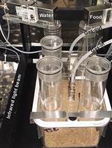
Testing Effects of Chronic Chemogenetic Neuronal Stimulation on Energy Balance by Indirect Calorimetry

Plant Science
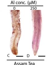
Qualitative Analysis of Lipid Peroxidation in Plants under Multiple Stress Through Schiff’s Reagent: A Histochemical Approach
.JPG)


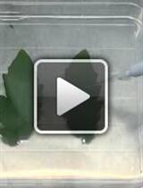
In-vitro and in-planta Botrytis cinerea Inoculation Assays for Tomato

Quantification of Thrips Damage Using Ilastik and ImageJ Fiji