
- Submit a Protocol
- Receive Our Alerts
- EN

- Protocols

- Articles and Issues
- For Authors
- About
- Become a Reviewer

Past Issue in 2018
Volume: 8, Issue: 10
Cancer Biology
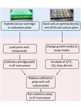
Measurement of Oxygen Consumption Rate (OCR) and Extracellular Acidification Rate (ECAR) in Culture Cells for Assessment of the Energy Metabolism

Cell Biology
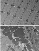
Transmission Electron Microscopy for Analysis of Mitochondria in Mouse Skeletal Muscle

Ex vivo Follicle Rupture and in situ Zymography in Drosophila

Intra-amniotic Injection of Mouse Embryos
Developmental Biology
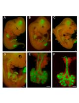
In vitro Explant Cultures to Interrogate Signaling Pathways that Regulate Mouse Lung Development
Immunology
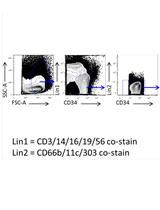
High Dimensional Functionomic Analysis of Human Hematopoietic Stem and Progenitor Cells at a Single Cell Level

Microbiology
Virucidal and Neutralizing Activity Tests for Antiviral Substances and Antibodies

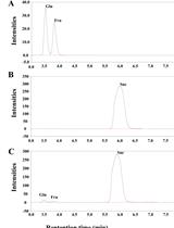
Enzymatic Activity Assay for Invertase in Synechocystis Cells
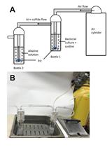
Quantification of Hydrogen Sulfide and Cysteine Excreted by Bacterial Cells
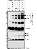
In vitro Analysis of Ubiquitin-like Protein Modification in Archaea

Molecular Biology
Ectopic Gene Expression in Macrophages Using in vitro Transcribed mRNA

Neuroscience
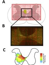
Induction of Photothrombotic Stroke in the Sensorimotor Cortex of Rats and Preparation of Tissue for Analysis of Stroke Volume and Topographical Cortical Localization of Ischemic Infarct
.jpg)
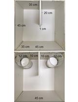
Sociability and Social Novelty Preference Tests Using a U-shaped Two-choice Field
In vivo Use of Dextran-based Anterograde Cortical Tracers to Assess the Integrity of the Cortical Spinal Tract
Plant Science
Quantification of Salicylic Acid (SA) and SA-glucosides in Arabidopsis thaliana


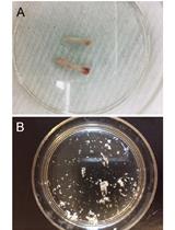
Rapid Screening and Evaluation of Maize Seedling Resistance to Stalk Rot Caused by Fusarium spp.
Stem Cell

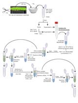
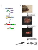
A Method to Injure, Dissect and Image Indirect Flight Muscle of Drosophila
Hair Follicle Stem Cell Isolation and Expansion
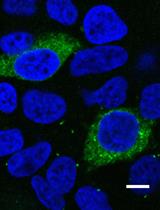
Osteoblast Sorting and Intracellular Staining of CXCL12
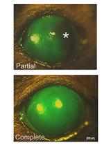
Murine Hair Follicle Derived Stem Cell Transplantation onto the Cornea Using a Fibrin Carrier