
- Submit a Protocol
- Receive Our Alerts
- EN

- Protocols

- Articles and Issues
- For Authors
- About
- Become a Reviewer

Past Issue in 2018
Volume: 8, Issue: 18
Biochemistry
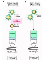
Retroviral Capsid Core Stability Assay


Cancer Biology

Zebrafish Embryo Xenograft and Metastasis Assay


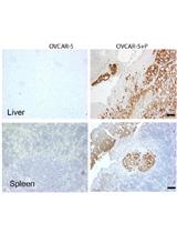
Qualitative in vivo Bioluminescence Imaging

Cell Biology
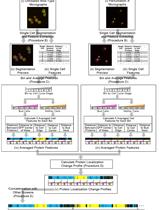
Extracting and Integrating Protein Localization Changes from Multiple Image Screens of Yeast Cells

Developmental Biology
Activation of Fibroblast Contractility via Cell-Cell Interactions and Soluble Signals


Immunology
Platelet Migration and Bacterial Trapping Assay under Flow
Microbiology
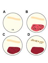
Plant Assays for Quantifying Ralstonia solanacearum Virulence

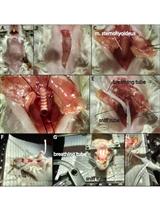
Sendai Virus Propagation Using Chicken Eggs
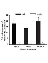
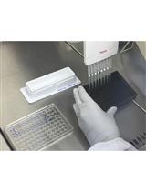
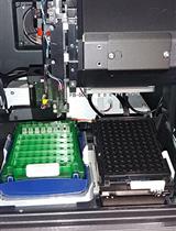
Extraction and Quantification of Polyphosphate (polyP) from Gram-negative Bacteria
High-throughput Microscopic Analysis of Salmonella Invasion of Host Cells


Molecular Biology
Identifying Protein Interactions with Histone Peptides Using Bio-layer Interferometry


Neuroscience
6-hydroxydopamine (6-OHDA) Oxidative Stress Assay for Observing Dopaminergic Neuron Loss in Caenorhabditis elegans
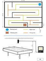
Classic Labyrinth Test for Neurobehavioral Evaluation in Wistar Rats

Assessing Classical Olfactory Fear Conditioning by Behavioral Freezing in Mice
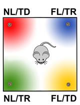
Behavioral Evaluation of Odor Memory in Mice
Artificial Inhalation Protocol in Adult Mice
A Behavioral Assay to Examine the Effects of Kavalactones on Caenorhabditis elegans Neuromuscular Excitability
Plant Science
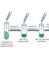
GC-MS-Based Analysis of Methanol: Chloroform-extracted Fatty Acids from Plant Tissues

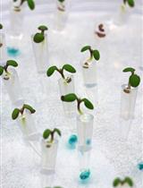
Fabrication and Use of the Dual-Flow-RootChip for the Imaging of Arabidopsis Roots in Asymmetric Microenvironments


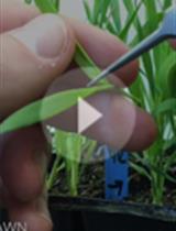
Live Confocal Imaging of Brachypodium Spikelet Meristems

Systems Biology
Stable-isotope Labeled Metabolic Analysis in Drosophila melanogaster: From Experimental Setup to Data Analysis