
- Submit a Protocol
- Receive Our Alerts
- EN

- Protocols

- Articles and Issues
- For Authors
- About
- Become a Reviewer

Past Issue in 2018
Volume: 8, Issue: 19
Biochemistry
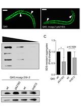
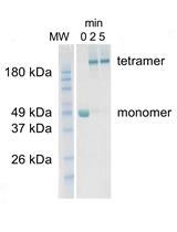
Filter Retardation Assay for Detecting and Quantifying Polyglutamine Aggregates Using Caenorhabditis elegans Lysates

A Method for SUMO Modification of Proteins in vitro
Preparation of Sequencing RNA Libraries through Chemical Cross-linking Coupled to Affinity Purification (cCLAP) in Saccharomyces cerevisiae

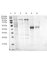
Expression and Ni-NTA-Agarose Purification of Recombinant Hepatitis C Virus E2 Ectodomain Produced in a Baculovirus Expression System
Cell Biology
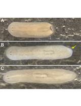
Maintenance of Schmidtea mediterranea in the Laboratory

Developmental Biology
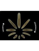
Detection of Cell Death in Planarians
Immunology

Isolation and Culture of Mouse Lung ILC2s


.JPG)
.JPG)
Microbiology
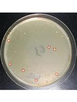
CRISPR/Cas9-mediated ssDNA Recombineering in Corynebacterium glutamicum


Molecular Biology
Fluorescence Titrations to Determine the Binding Affinity of Cyclic Nucleotides to SthK Ion Channels


Neuroscience
Isolation of Chromatin-bound Proteins from Subcellular Fractions for Biochemical Analysis

Measurement of Dopamine Using Fast Scan Cyclic Voltammetry in Rodent Brain Slices

Drosophila Endurance Training and Assessment of Its Effects on Systemic Adaptations
Plant Science
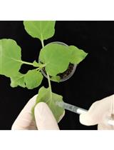
Real-time PCR Analysis of PAMP-induced Marker Gene Expression in Nicotiana benthamiana

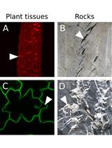
An Image Analysis Pipeline to Quantify Emerging Cracks in Materials or Adhesion Defects in Living Tissues
