
- Submit a Protocol
- Receive Our Alerts
- EN

- Protocols

- Articles and Issues
- For Authors
- About
- Become a Reviewer

Past Issue in 2018
Volume: 8, Issue: 20
Biochemistry
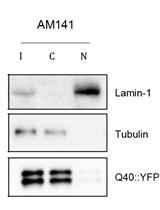
Nuclear/Cytoplasmic Fractionation of Proteins from Caenorhabditis elegans

Cell Biology

Murine Pharmacokinetic Studies


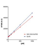
Transcytosis Assay for Transport of Glycosphingolipids across MDCK-II Cells


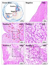
Vascular Permeability Assay in Human Coronary and Mouse Brachiocephalic Arteries
Immunology
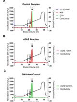
A Highly Sensitive Anion Exchange Chromatography Method for Measuring cGAS Activity in vitro
Microbiology
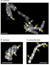
Assessing Membrane Fluidity and Visualizing Fluid Membrane Domains in Bacteria Using Fluorescent Membrane Dyes

HIVGKO: A Tool to Assess HIV-1 Latency Reversal Agents in Human Primary CD4+ T Cells
Preparation and Purification of Proteins Secreted from Phytophthora sojae

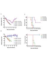
H1N1 Virus Production and Infection

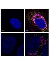
Detection and Differentiation of Multiple Viral RNAs Using Branched DNA FISH Coupled to Confocal Microscopy and Flow Cytometry
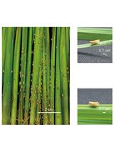
Rice Ragged Stunt Virus Propagation and Infection on Rice Plants

Molecular Biology
Generation of Gene Knockout and Gene Replacement with Complete Removal of Full-length Endogenous Transcript Using CRISPR-Trap


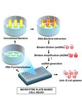
Microtitre Plate Based Cell-SELEX Method
Neuroscience
Optical Clearing and Index Matching of Tissue Samples for High-resolution Fluorescence Imaging Using SeeDB2
Behavioral Evaluation of Seeking and Preference of Alcohol in Mice Subjected to Stress

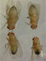
Testing for Assortative Mating by Diet in Drosophila melanogaster
Plant Science
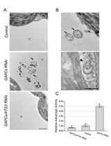
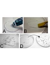
Examining Autophagy in Plant by Transmission Electron Microscopy (TEM)


Pneumatic Method to Measure Plant Xylem Embolism