
- Submit a Protocol
- Receive Our Alerts
- EN

- Protocols

- Articles and Issues
- For Authors
- About
- Become a Reviewer

Past Issue in 2018
Volume: 8, Issue: 23

Biochemistry
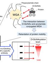
Wheat Germ Agglutinin (WGA)-SDS-PAGE: A Novel Method for the Detection of O-GlcNAc-modified Proteins by Lectin Affinity Gel Electrophoresis
Purification of Globular Actin from Rabbit Muscle and Pyrene Fluorescent Assays to Investigate Actin Dynamics in vitro
In vitro Membrane Interaction and Liposome Fusion Assays Using Recombinant Hepatitis C Virus Envelope Protein E2
Cancer Biology
Cluster Analysis of Endogenous HER2 and HER3 Receptors in SKBR3 Cells

Cell Biology
Blinded Visual Scoring of Images Using the Freely-available Software Blinder
Developmental Biology

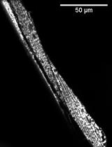
Notochord Injury Assays that Stimulate Transcriptional Responses in Zebrafish Larvae

Immunology
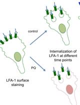
Flow Cytometry Assay for Recycling of LFA-1 in T-lymphocytes
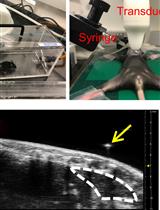
Ultrasound Guided Intra-thymic Injection to Track Recent Thymic Emigrants and Investigate T Cell Development
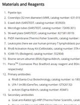
Integrin Dependent RhoB Activation Assay Using Leukocytes
Microbiology
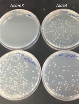
Quantitative Transformation Efficiency Assay for Bacillus subtilis
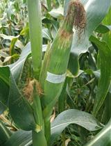
Field Inoculation and Classification of Maize Ear Rot Caused by Fusarium verticillioides


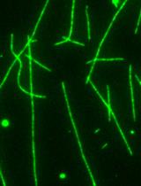
An in vitro Microscopy-based Assay for Microtubule-binding and Microtubule-crosslinking by Budding Yeast Microtubule-associated Protein

Plant Science
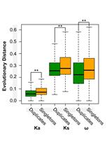
Genome-wide Estimation of Evolutionary Distance and Phylogenetic Analysis of Homologous Genes

Using Arabidopsis Mesophyll Protoplasts to Study Unfolded Protein Response Signaling
