
- Submit a Protocol
- Receive Our Alerts
- EN

- Protocols

- Articles and Issues
- For Authors
- About
- Become a Reviewer

Past Issue in 2015
Volume: 5, Issue: 10
Cancer Biology

Proximity Ligation Assay (PLA) to Detect Protein-protein Interactions in Breast Cancer Cells


Genome-Wide siRNA Screen for Anti-Cancer Drug Resistance in Adherent Cell Lines
Immunology
Murine Liver Myeloid Cell Isolation Protocol

Isolation of Particles of Recombinant ASC and NLRP3
Microbiology
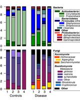
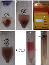
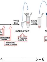
Human, Bacterial and Fungal Amplicon Collection and Processing for Sequencing
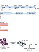
Determining the Relative Fitness Score of Mutant Viruses in a Population Using Illumina Paired-end Sequencing and Regression Analysis
Molecular Biology
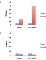
RNA-binding Protein Immunoprecipitation (RIP) to Examine AUF1 Binding to Senescence-Associated Secretory Phenotype (SASP) Factor mRNA
Plant Science
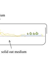
Establishment of a Symbiotic in vitro System between a Green Meadow Orchid and a Rhizoctonia-like Fungus
Plant Materials and Growth Conditions of Japanese Morning Glory (Ipomoea nil cv. Violet)

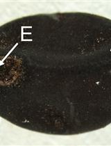
Histochemical Detection of Zn in Plant Tissues


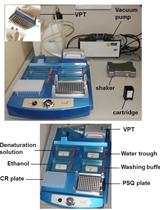
Pyrosequencing Approach for SNP Genotyping in Plants Using a M13 Biotinylated Primer


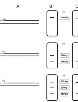
Fluorescence-based CAPS Multiplex Genotyping on Capillary Electrophoresis Systems

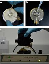
Measurement of Mitochondrial Respiration Rate in Maize (Zea mays) Leaves


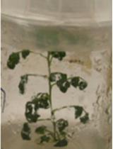
Transient Transformation of Artemisia annua

