
- Submit a Protocol
- Receive Our Alerts
- EN

- Protocols

- Articles and Issues
- For Authors
- About
- Become a Reviewer

Past Issue in 2019
Volume: 9, Issue: 8
Biochemistry
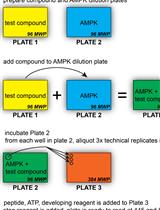
Non-radiometric Cell-free Assay to Measure the Effect of Molecular Chaperones on AMP-activated Kinase Activity

Cancer Biology
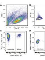
Measuring Protein Synthesis during Cell Cycle by Azidohomoalanine (AHA) Labeling and Flow Cytometric Analysis
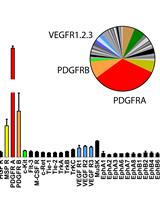
Unbiased Screening of Activated Receptor Tyrosine Kinases (RTKs) in Tumor Extracts Using a Mouse Phospho-RTK Array Kit
Cell Biology
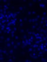
Immunofluorescence Staining of WT-1/Podocalyxin on Mouse Kidney Sections

Immunology
Evaluation of Mucosal and Systemic Vaccine Responses by Cyclic di-GMP (CDG)-adjuvanted Protein Subunit Vaccines

Molecular Biology
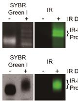
Northern Blot with IR Fluorescent Probes: Strategies for Probe Preparation


Neuroscience
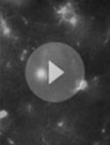
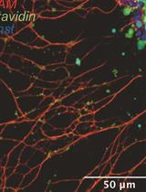
Time-lapse Whole-field Fluorescence Imaging of Microglia Processes Motility in Acute Mouse Hippocampal Slices and Analysis


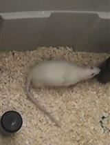
Displaced Object Recognition Memory in Rats

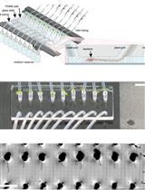
Preparation and Manipulation of Olfactory Epithelium Explant Cultures for Measurement of the Mechanical Tension of Individual Axons Using the Biomembrane Force Probe
Plant Science
Tracking Root Interactions System (TRIS) Experiment and Quality Control