
- Submit a Protocol
- Receive Our Alerts
- EN

- Protocols

- Articles and Issues
- For Authors
- About
- Become a Reviewer

Past Issue in 2019
Volume: 9, Issue: 17
Biochemistry
![Photoaffinity Labeling of Respiratory Complex I in Bovine Heart Submitochondrial Particles by Photoreactive [125I] amilorides](https://en-cdn.bio-protocol.org/imageup/arcimg/20190902091054730.jpg?t=1758522982)
Photoaffinity Labeling of Respiratory Complex I in Bovine Heart Submitochondrial Particles by Photoreactive [125I] amilorides
Cancer Biology
A Robust, One-step FRET Assay for Human Heparanase
Cell Biology
In vitro Intestinal Epithelial Wound-healing Assays Using Electric Cell-Substrate Impedance Sensing Instrument

Developmental Biology
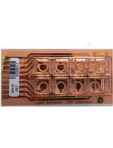
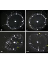
Estrous Cycle Monitoring in Mice with Rapid Data Visualization and Analysis

A Widely Applicable Urea-based Fluorescent/Colorimetric mRNA in situ Hybridization Protocol
Immunology
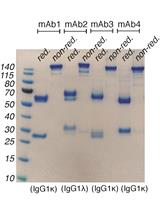
A Refined Protocol for Identifying Citrulline-specific Monoclonal Antibodies from Single Human B Cells from Rheumatoid Arthritis Patient Material
Isolation of Neutrophil Nuclei for Use in NETosis Assays
Microbiology
Yeast Single-cell RNA-seq, Cell by Cell and Step by Step
High Resolution Respirometry in Candida albicans
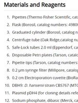
Development of an Efficient Transformation System for Halotolerant Yeast Debaryomyces hansenii CBS767
Neuroscience
Co-culture of Human Stem Cell Derived Neurons and Oligodendrocyte Progenitor Cells
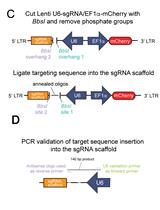
A Novel Dual Lentiviral CRISPR-based Transcriptional Activation System for Gene Expression Regulation in Neurons
A High-throughput and Pathophysiologically Relevant Astrocyte-motor Neuron Co-culture Assay for Amyotrophic Lateral Sclerosis Therapeutic Discovery
Plant Science
PEG-mediated, Stable, Nuclear and Chloroplast Transformation of Cyanidioschizon merolae
