
- Submit a Protocol
- Receive Our Alerts
- EN

- Protocols

- Articles and Issues
- For Authors
- About
- Become a Reviewer

Past Issue in 2020
Volume: 10, Issue: 1
Biophysics
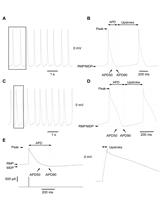
Whole-cell and Perforated Patch-clamp Recordings from Acutely-isolated Murine Sino-atrial Node Cells
Cancer Biology
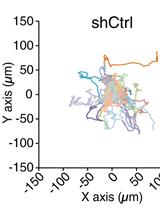
Analysis of Random Migration of Cancer Cells in 3D


Cell Biology
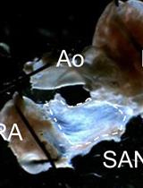
Acute Isolation of Cells from Murine Sino-atrial Node
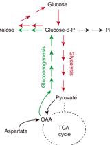
Steady-state and Flux-based Trehalose Estimation as an Indicator of Carbon Flow from Gluconeogenesis or Glycolysis

Developmental Biology
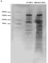
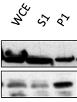
RNA Interactome Identification via RNA-BioID in Mouse Embryonic Fibroblasts


Microbiology
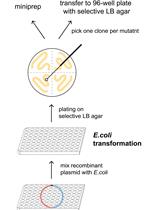
Unbiased and Tailored CRISPR/Cas gRNA Libraries by Synthesizing Covalently-closed-circular (3Cs) DNA
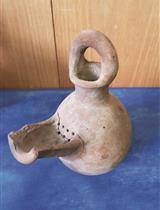
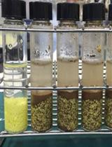
Isolation and Characterization of Live Yeast Cells from Ancient Clay Vessels


A Microbial Bioassay for Direct Contact Assessment of Soil Toxicity Based on Oxygen Consumption of Sulfur Oxidizing Bacteria
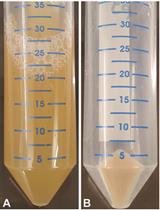
A Yeast Chromatin-enriched Fractions Purification Approach, yChEFs, from Saccharomyces cerevisiae

Molecular Biology
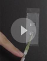
High-throughput Site-directed Scanning Mutagenesis Using a Two-fragment PCR Approach


Neuroscience
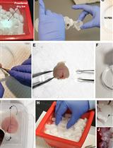
Laser Capture Micro-dissection (LCM) of Neonatal Mouse Forebrain for RNA Isolation


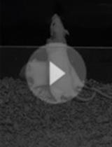
Assessing Rough-and-tumble Play Behavior in Juvenile Rats

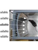
The Mouse Gambling Task: Assessing Individual Decision-making Strategies in Mice
Plant Science
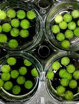
Visualization of Actin Organization and Quantification in Fixed Arabidopsis Pollen Grains and Tubes


Measurement of Chloroplastic NAD Kinase Activity and Whole Tissue NAD Kinase Assay