
- Submit a Protocol
- Receive Our Alerts
- EN

- Protocols

- Articles and Issues
- For Authors
- About
- Become a Reviewer

Past Issue in 2020
Volume: 10, Issue: 11
Biochemistry
Biochemical Pulldown of mRNAs and Long Noncoding RNAs from Cellular Lysates Coupled with Mass Spectrometry to Identify Protein Binding Partners


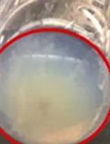
Isolating Multiple Extracellular Vesicles Subsets, Including Exosomes and Membrane Vesicles, from Bovine Milk Using Sodium Citrate and Differential Ultracentrifugation

Cell Biology
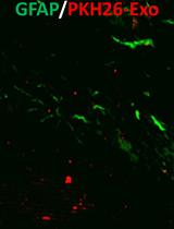
Extracellular Vesicles Tracking and Quantification Using CT and Optical Imaging in Rats


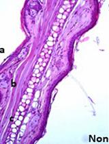
Ex vivo Culture Assay Using Human Hair Follicles to Study Circadian Characteristics
Developmental Biology

FRET Reporter Assays for cAMP and Calcium in a 96-well Format Using Genetically Encoded Biosensors Expressed in Living Cells


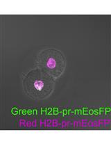
Primed Track: Reliable Volumetric Single-cell Tracking and Lineage Tracing of Living Specimen with Dual-labeling Approaches

Immunology
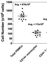
Methodology for in vitro Assessment of Human T Cell Activation and Blockade
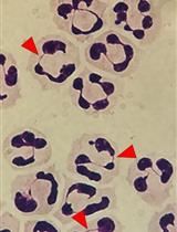
Isolation and High Throughput Flow Cytometric Apoptosis Assay of Human Neutrophils to Enable Compound Library Screening
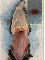
RNA Extraction from Ears and Draining Lymph Nodes of Mice Infected with Leishmania amazonensis

Microbiology
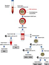
Differential Fractionation of Erythrocytes Infected by Plasmodium berghei
Molecular Biology
Mouse Adipose Tissue Protein Extraction
Neuroscience
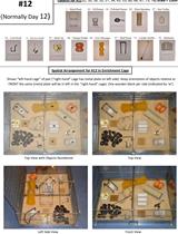
Enriched Environment Procedures for Rodents: Creating a Standardized Protocol for Diverse Enrichment to Improve Consistency across Research Studies

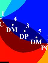
Precise Targeting of Single Microelectrodes to Orientation Pinwheel Centers
Plant Science
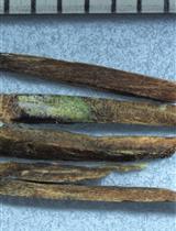
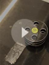
Safe DNA-extraction Protocol Suitable for Studying Tree-fungus Interactions
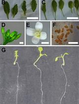
Determination of Ureides Content in Plant Tissues
