
- Submit a Protocol
- Receive Our Alerts
- EN

- Protocols

- Articles and Issues
- For Authors
- About
- Become a Reviewer

Past Issue in 2020
Volume: 10, Issue: 12

Biochemistry
Electrophoretic Mobility Shift Assay of in vitro Phosphorylated RNA Polymerase II Carboxyl-terminal Domain Substrates
Preparation of HeLa Total Membranes and Assay of Lipid-inhibition of Serine Palmitoyltransferase Activity
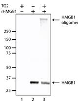
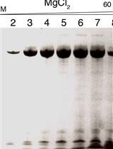
In vitro Crosslinking Reactions and Substrate Incorporation Assays for The Identification of Transglutaminase-2 Protein Substrates

Biophysics
Preparation of Yeast tRNA Sample for NMR Spectroscopy

Cell Biology
Superresolution Microscopy of Drosophila Indirect Flight Muscle Sarcomeres


Quantification of Protein Kinase A (PKA) Activity by An in vitro Radioactive Assay Using the Mouse Sperm Derived Enzyme


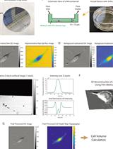
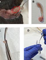
Single Cell Volume Measurement Utilizing the Fluorescence Exclusion Method (FXm)

Immunology
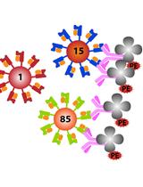
Assessments of HLA-I Specificities of Anti-HLA-I Monoclonal Antibodies Using Solid Phase Bead Arrays
Microbiology
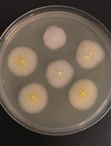
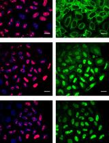
Genomic Edition of Ashbya gossypii Using One-vector CRISPR/Cas9
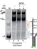
Site-specific DNA Mapping of Protein Binding Orientation Using Azidophenacyl Bromide (APB)
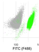
Live Cell Measurement of the Intracellular pH of Yeast by Flow Cytometry Using a Genetically-Encoded Fluorescent Reporter
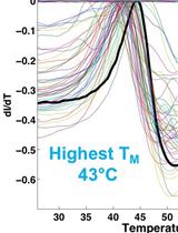
Identification of Buffer Conditions for Optimal Thermostability and Solubility of Herpesviral Protein UL37 Using the Thermofluor Assay

Neuroscience

An Alternative Maze to Assess Novel Object Recognition in Mice
Quantitative Nucleocytoplasmic Transport Assays in Cellular Models of Neurodegeneration
Plant Science
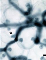
Maintenance and Quantitative Phenotyping of the Oomycete-plant Model Pathosystem Hyaloperonospora arabidopsidis–Arabidopsis

Systems Biology

Low-cost and Multiplexable Whole mRNA-Seq Library Preparation Method with Oligo-dT Magnetic Beads for Illumina Sequencing Platforms