
- Submit a Protocol
- Receive Our Alerts
- EN

- Protocols

- Articles and Issues
- For Authors
- About
- Become a Reviewer

Past Issue in 2020
Volume: 10, Issue: 24
Biochemistry
Preparation of Bacterial Outer Membrane Vesicles for Characterisation of Periplasmic Proteins in Their Native Environment

Triacylglycerol Measurement in HeLa Cells

Quantitative Irreversible Tethering (qIT) for Target-directed Covalent Fragment Screening


Cancer Biology
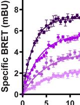
Equilibrium and Kinetic Measurements of Ligand Binding to HiBiT-tagged GPCRs on the Surface of Living Cells


Developmental Biology
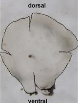
Whole-mount Immunohistochemistry of Adult Zebrafish Retina for Advanced Imaging

Immunology
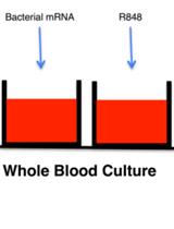
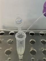
RNA ImmunoGenic Assay: A Method to Detect Immunogenicity of in vitro Transcribed mRNA in Human Whole Blood

Pea Aphid Rearing, Bacterial Infection and Hemocyte Phagocytosis Assay

Microbiology
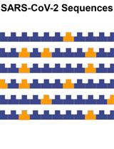
Evaluation of the Sequence Variability within the PCR Primer/Probe Target Regions of the SARS-CoV-2 Genome
Antimicrobial Sensitivity Assay for Bdellovibrio bacteriovorus
Preparation of Nippostrongylus brasiliensis Larvae for the Study of Host Skin Response

Neuroscience
A Quantitative Assay to Measure Stress Granule Association of Proteins and Peptides in Semi-permeabilized Human Cells
Nestlet Shredding and Nest Building Tests to Assess Features of Psychiatric Disorders in Mice


Plant Science
Lipid Droplet Isolation from Arabidopsis thaliana Leaves
![Analysis of Isotopically-labeled Monogalactosyldiacylglycerol Molecular Species from [14C]Acetate-Labeled Tobacco Leaves](https://en-cdn.bio-protocol.org/imageup/arcimg/20201215013528186.jpg?t=1758512010)
Analysis of Isotopically-labeled Monogalactosyldiacylglycerol Molecular Species from [14C]Acetate-Labeled Tobacco Leaves

Stem Cell

Isolation of Extracellular Vesicles Derived from Mesenchymal Stromal Cells by Ultracentrifugation


Differentiation of Human Induced Pluripotent Stem Cells (hiPSCs) into Osteoclasts