
- Submit a Protocol
- Receive Our Alerts
- EN

- Protocols

- Articles and Issues
- For Authors
- About
- Become a Reviewer

Past Issue in 2021
Volume: 11, Issue: 2
Biochemistry
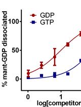
A mant-GDP Dissociation Assay to Compare the Guanine Nucleotide Binding Preference of Small GTPases

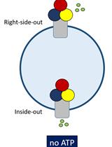
Polyamine Transport Assay Using Reconstituted Yeast Membranes

Reverse Phase-high-performance Liquid Chromatography (RP-HPLC) Analysis of Globin Chains from Human Erythroid Cells

Cancer Biology
Evaluation of Toxicity with Brine Shrimp Assay

Imaging of Human Cancer Cells in 3D Collagen Matrices
Immunology
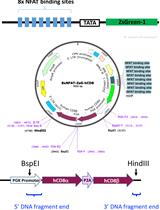
Multiplex T-cell Stimulation Assay Utilizing a T-cell Activation Reporter-based Detection System
Wounding Caenorhabditis elegans with Glass Wool


Microbiology
A One-enzyme RT-qPCR Assay for SARS-CoV-2, and Procedures for Reagent Production
Analysis of Pseudomonas aeruginosa c-di-GMP High and Low Subpopulations Using Flow-assisted Cell Sorting (FACS) and Quantitative Reverse Transcriptase PCR (qRT-PCR)
Determination of Antibacterial Activity of Film Coatings against Four Clinically Relevant Bacterial Strains
Bacterial Conjugation Protocol for Ruminant Mycoplasmas
Bacterial Adhesion Kinetics in a High Throughput Setting in Seconds-minutes Time Resolution

Neuroscience
Preparation of Synaptoneurosomes to Study the Synapse in the Murine Cerebral Cortex
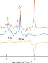
Low- and High-resolution Dynamic Analyses for Magnetic Resonance Spectroscopy Data
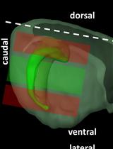
Characterization of Hippocampal Adult-borne Granule Cells in a Transient Cerebral Ischemia Model
Plant Science
Cellulase and Macerozyme-PEG-mediated Transformation of Moss Protoplasts


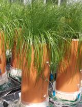
Design and Construction of Repacked Soil Columns for Measuring Solute Transport, Plant Growth and Soil Biota
.jpg)
Stem Cell
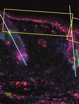
Histological and Immunohistochemical Examination of Stem Cell Proliferation and Reepithelialization in the Wounded Skin

