
- Submit a Protocol
- Receive Our Alerts
- EN

- Protocols

- Articles and Issues
- For Authors
- About
- Become a Reviewer

Past Issue in 2021
Volume: 11, Issue: 13
Biochemistry

Visualization of Host Cell Kinase Activation by Viral Proteins Using GFP Fluorescence Complementation and Immunofluorescence Microscopy
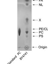
Yeast Lipid Extraction and Analysis by HPTLC
Biological Engineering
Building a Total Internal Reflection Microscope (TIRF) with Active Stabilization (Feedback SMLM)
Biophysics
OrganoPlate Micro-fluidic Microvessel Culture and Analysis
Single-molecule Fluorescence Technique to Monitor the Co-transcriptional Formation of G-quadruplex and R-loop Structures
Cancer Biology
Monitoring Changes in the Oxidizing Milieu in the Endoplasmic Reticulum of Mammalian Cells Using HyPerER
Cell Biology

A Method for Estimating the Potential Synaptic Connections Between Axons and Dendrites From 2D Neuronal Images
Electron Tomography to Study the Three-dimensional Structure of the Reovirus Egress Pathway in Mammalian Cells

Ethanol-induced Sedative Behavior: An Assay to Investigate Increased Dopamine Signaling in Caenorhabditis elegans
Microbiology
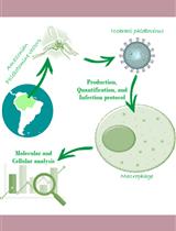
Production, quantification, and infection of Amazonian Phlebovirus (Bunyaviridae)
Molecular Biology
Measurement of Transgene Copy Number in Plants Using Droplet Digital PCR
PCR-mediated One-day Synthesis of Guide RNA for the CRISPR/Cas9 System
Neuroscience

Immunofluorescence of GFAP and TNF-α in the Mouse Hypothalamus
An Improved Method for Individual Tracking of Voluntary Wheel Running in Pair-housed Juvenile Mice
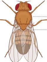
Evaluating Baseline and Sensitised Heat Nociception in Adult Drosophila
Plant Science
Analysis of Soluble Sugar Content in Minute Quantities of Rice Tissues by GC-MS