
- Submit a Protocol
- Receive Our Alerts
- EN

- Protocols

- Articles and Issues
- For Authors
- About
- Become a Reviewer

Past Issue in 2015
Volume: 5, Issue: 16
Biochemistry
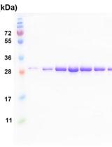
Sample Preparation of Telomerase Subunits for Crystallization

Cancer Biology
Determining Leukocyte Origins Using Parabiosis in the PyMT Breast Tumor Model

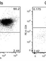
Adoptive Transfer of Myeloid-Derived Suppressor Cells and T Cells in a Prostate Cancer Model

Cell Biology
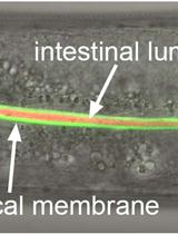
Labeling of the Intestinal Lumen of Caenorhabditis elegans by Texas Red-dextran Feeding

Immunology
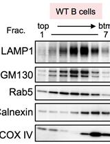
Separation of Intracellular Vesicles for Immunoassays
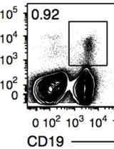
Adoptive Transfer of Memory B Cells

Microbiology
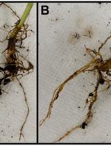
Isolation of Rhizosphere Bacterial Communities from Soil


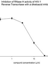
RNase H Polymerase-independent Cleavage Assay for Evaluation of RNase H Activity of Reverse Transcriptase Enzymes

Plant Science
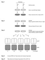
Protocol to Treat Seedlings with Brassinazole and Measure Hypocotyl Length in Arabidopsis thaliana

In vitro Colorimetric Method to Measure Plant Glutamate Dehydrogenase Enzyme Activity


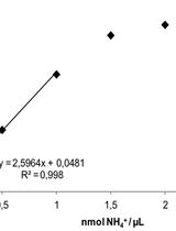
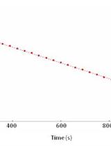
High-throughput Quantification of Ammonium Content in Arabidopsis
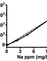
Quantification of Sodium Accumulation in Arabidopsis thaliana Using Inductively Coupled Plasma Optical Emission Spectrometery (ICP-OES)


Glucosinolates Determination in Tissues of Horseradish Plant


Stem Cell

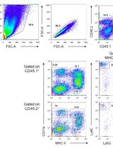
FACS-based Satellite Cell Isolation From Mouse Hind Limb Muscles


Systems Biology
Character-State Reconstruction to Infer Ancestral Protein-Protein Interaction Patterns
