
- Submit a Protocol
- Receive Our Alerts
- EN

- Protocols

- Articles and Issues
- For Authors
- About
- Become a Reviewer

Past Issue in 2015
Volume: 5, Issue: 19
Cell Biology
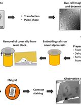
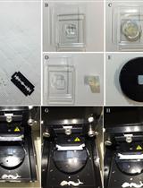
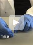
Sample Preparation for Correlative Light and Electron Microscopy (CLEM) Analyses in Cellular Microbiology
Immunology
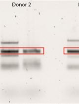
TCRβ Clonotype Analysis of EBV and CMV-specific Human CD8+ T Cells
Microbiology
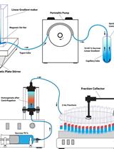
Density Gradient Centrifugation for Enrichment and Identification of GFP-tagged Chitosomal Microvesicles of Filamentous Fungi

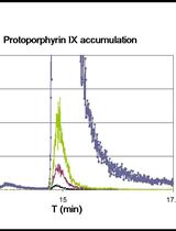
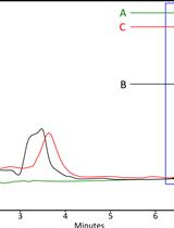
Bacterial Porphyrin Extraction and Quantification by LC/MS/MS Analysis
In vitro Studies: Inhibition of Nevirapine Metabolism by Nortriptyline in Hepatic Microsomes


Neuroscience

Chick Neural Tube Explant Culture


Plant Science
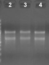
3’ Rapid Amplification of cDNA Ends (3’ RACE) Using Arabidopsis Samples


Quantification of Callose Deposition in Plant Leaves


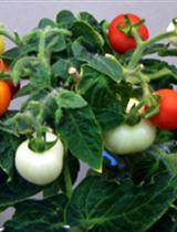
Hydroponic Culture of ‘Micro-Tom’ Tomato

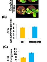
Pot Level Drought Stress Tolerance Assay in Tobacco through Plant Phenotyping and Antioxidant Assay


Histochemical Staining of Silica Body in Rice Leaf Blades
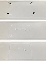
Pectin Nanostructure Visualization by Atomic Force Microscopy
Analysis of in vivo Cellulose Biosynthesis in Arabidopsis Cells by Spinning Disk Confocal Microscopy