
- Submit a Protocol
- Receive Our Alerts
- EN

- Protocols

- Articles and Issues
- For Authors
- About
- Become a Reviewer

Past Issue in 2015
Volume: 5, Issue: 20
Cancer Biology
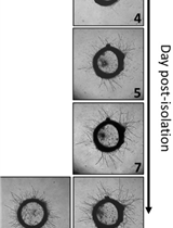
Rat Aortic Ring Model to Assay Angiogenesis ex vivo


Cell Biology
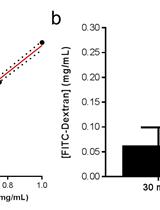
In vivo Fluorescein Isothiocyanate-dextran (FD4) Permeability Assay
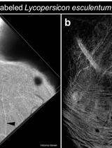
Intravenous Tomato Lectin Injection to Assess Functional Vasculature
Immunology
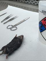
Immune Cell Isolation from Mouse Femur Bone Marrow

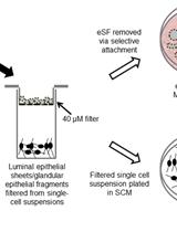
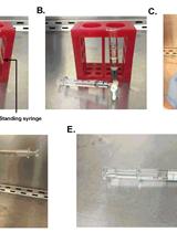
Isolation and Culture of Human Endometrial Epithelial Cells and Stromal Fibroblasts

Collagen-induced Arthritis: A Model for Murine Autoimmune Arthritis


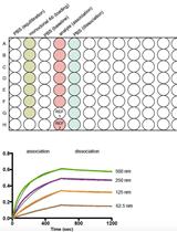
Kinetic Analysis of Monoclonal Antibody Binding to HIV-1 gp120-derived Hyperglycosylated Cores
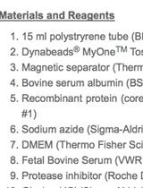
Coupling of HIV-1 gp120-derived Core Protein to Paramagnetic Beads and Adsorption Assays
Microbiology
Product Analysis of Starch Active Enzymes by TLC

Purification of a Protein Exhibiting Isoleucine 2-epimerase Activity from Lactobacillus otakiensis JCM 15040
Plant Science
Hyaloperonospora arabidopsidis (Downy Mildew) Infection Assay in Arabidopsis


Isolation of Triterpenes from Propolis (Bee Glue)
Isolation and Characterization Procedure for Indole Alkaloids from the Marquesan Plant Rauvolfia Nukuhivensis

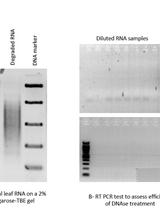
Mitochondrial RNA Transcript Analysis Assay of Arabidopsis Leaf Tissues
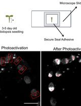
Two-photon Photoactivation to Measure Histone Exchange Dynamics in Plant Root Cells
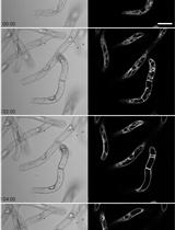
Vacuole Structure Analysis during Cell Death Subsequent to Application of Erwinia carotovora Culture Filtrates to Cell Cultures of Nicotiana tabacum


Stem Cell
Clonal Culture of Mouse Liver Progenitor Cells