
- Submit a Protocol
- Receive Our Alerts
- EN

- Protocols

- Articles and Issues
- For Authors
- About
- Become a Reviewer

Past Issue in 2015
Volume: 5, Issue: 22
Cancer Biology
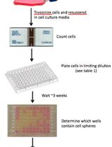
In vitro and in vivo Limiting Dilution Assay for Colorectal Cancer
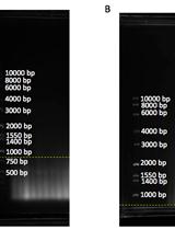
Telomere Restriction Fragment (TRF) Analysis


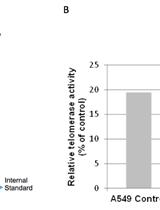
Telomerase Repeated Amplification Protocol (TRAP)
13C Tracer Studies of Metabolism in Mouse Tumor Xenografts
Telomere Dysfunction Induced Foci (TIF) Analysis
Immunology

Skin Wound Healing Model - Excisional Wounding and Assessment of Lesion Area


Estimation of Wound Tissue Neutrophil and Macrophage Accumulation by Measuring Myeloperoxidase (MPO) and N-Acetyl-β-D-glucosaminidase (NAG) Activities
Permanent Occlusion of the Left Anterior Coronary Artery in the Rat

Microbiology
Purification of Bacterial RNA from Infected Macrophages
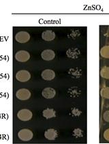
An Assay to Test the Capacity of Arabidopsis Plant Defensin Type1 Protein to Induce Cellular Zinc (Zn) Tolerance in Yeast
Neuroscience
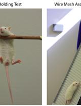
Locomotor Coordination Assay in Rats

Plant Science
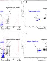
Sample Preparation and Fractionation of Arabidopsis thaliana Sperm and Vegetative Cell Nuclei by FACS

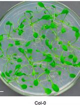
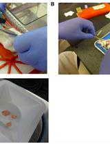
Arabidopsis Leaf Explant Culture


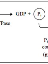
Expression, Purification and in vitro Enzyme Activity Assay of Plant Derived GTPase
Quantifying the Permeability of the Apoplastic Water Barrier in Cosmos Petals