
- Submit a Protocol
- Receive Our Alerts
- EN

- Protocols

- Articles and Issues
- For Authors
- About
- Become a Reviewer

Past Issue in 2015
Volume: 5, Issue: 23
Cancer Biology
Human Blood Component Vaccinia Virus Neutralization Assay


Cell Biology
Single Molecule RNA FISH in the Mammalian Oocyte

Immunology
Isolation of Lymphocytes from Murine Visceral Adipose Tissue


Excision of Visceral Adipose Tissue from Live Mice (VATectomy)

Microbiology
Establishing a Biofilm Co-culture of Pseudomonas and Aspergillus for Metabolite Extraction
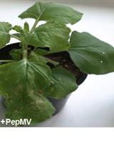
The Application of Quercetin to Study the Effect of Hsp70 Silencing on Plant Virus Infection in Nicotiana benthamiana Plants


Plant Science
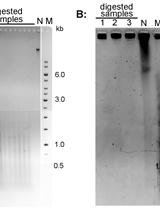
Terminal Restriction Fragments (TRF) Method to Analyze Telomere Lengths


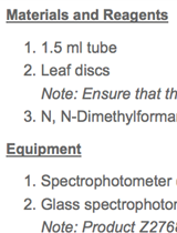
Purification of Rubisco from Chlamydomonas reinhardtii


Quantification of the Volume and Surface Area of Symbiosomes and Vacuoles of Infected Cells in Root Nodules of Medicago truncatula

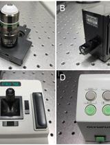
Analysis of Developing Pollen Grains within Intact Arabidopsis thaliana Anthers by Olympus Two-Photon Laser Scanning Microscopy


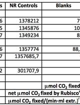
Assay of the Carboxylase Activity of Rubisco from Chlamydomonas reinhardtii
Chlorophyll Content Assay to Quantify the Level of Necrosis Induced by Different R Gene/Elicitor Combinations after Transient Expression
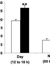
Measurement of H+ Flux in Rice by Non-invasive Micro-test Technology
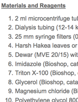
Extraction of Intracellular and Cell Wall Proteins from Leaves and Roots of Harsh Hakea

Rhizosphere Acidification Assay
