
- Submit a Protocol
- Receive Our Alerts
- EN

- Protocols

- Articles and Issues
- For Authors
- About
- Become a Reviewer

Past Issue in 2024
Volume: 14, Issue: 20
Cancer Biology

Optical Modulation of the Blood-Brain Barrier for Glioblastoma Treatment
Cell Biology
Purification and Immunostaining of Mouse Ependymal Ciliary Shafts

Developmental Biology
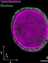
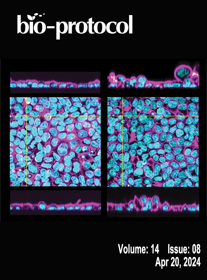
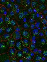
Fluorescence Imaging of 3D Cell Models with Subcellular Resolution
Immunology
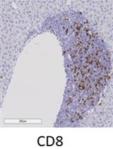
Immunohistochemistry of Immune Cells and Cells Bound to in vivo Administered Antibodies in Liver, Lung, Pancreas, and Colon of B6/lpr Mice
Microbiology
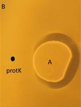
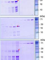
Genome-assisted Identification, Purification, and Characterization of Bacteriocins
Binding Affinity Quantifications of the Bacteriophage Mu DNA Modification Protein Mom Using Microscale Thermophoresis (MST)
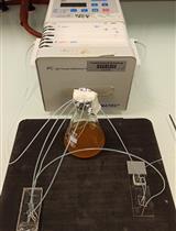
A Microfluidic Platform for Tracking Individual Cell Dynamics during an Unperturbed Nutrients Exhaustion
Neuroscience
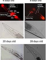
Analysis of Caenorhabditis elegans Aging-related Neurodegeneration in Chemosensory Neurons
Maximizing the Rod Outer Segment Yield in Retinas Extracted from Cattle Eyes
Stem Cell
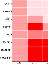
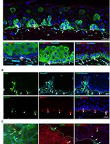
Gene Expression Analysis in Stem Cell-derived Cortical Neuronal Cultures Using Multi-well SYBR Green Quantitative PCR Arrays
Isolation and ex vivo Expansion of Limbal Mesenchymal Stromal Cells