
- Submit a Protocol
- Receive Our Alerts
- EN

- Protocols

- Articles and Issues
- For Authors
- About
- Become a Reviewer

Past Issue in 2016
Volume: 6, Issue: 2
Cancer Biology
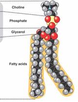
A Technique for the Measurement of in vitro Phospholipid Synthesis via Radioactive Labeling


Microbiology
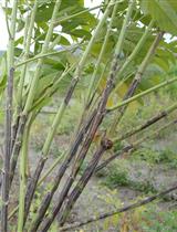
A Reliable Method for Phytophthora cajani Isolation, Sporangia, Zoospore Production and in Planta Infection of Pigeonpea


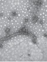
Preparation of Outer Membrane Vesicles from Myxococcus xanthus

HBV Infection in Human Hepatocytes and Quantification of Encapsidated HBV DNA

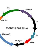
Infection of Human Hepatocyte-chimeric Mice with HBV and in vivo Treatment with εRNA
Molecular Biology
Identification of RNA-binding Proteins by RNA Ligand-based cDNA Expression Library Screening


Plant Science
Cotyledon Wounding of Arabidopsis Seedlings
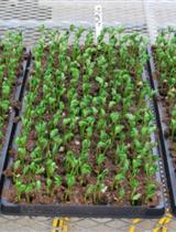
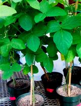
Agrobacterium rhizogenes-Based Transformation of Soybean Roots to Form Composite Plants


Soybean Cyst Nematode, Heterodera glycines, Infection Assay Using Soybean Roots
Analysis of Starch Synthase Activities in Wheat Grains using Native-PAGE

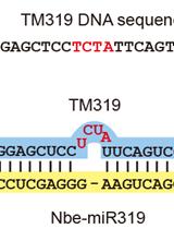
Virus-based MicroRNA Silencing


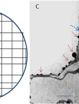
Cytohistochemical Determination of Calcium Deposition in Plant Cells

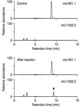
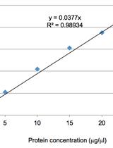
LC/MS-based Detection of Hydroxyproline O-galactosyltransferase Activity

Detection of Hydroxyproline O-galactoside by LC/MS