
- Submit a Protocol
- Receive Our Alerts
- EN

- Protocols

- Articles and Issues
- For Authors
- About
- Become a Reviewer

Past Issue in 2016
Volume: 6, Issue: 3
Cancer Biology
Stable Isotope Resolved Metabolomics Studies in ex vivo TIssue Slices
Cell Biology
Mouse Oocyte Isolation, Cultivation and RNA Microinjection


Immunology
Mouse BMDC-dependent T Cell Polarization Assays

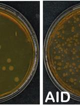
Cell-based Assays to Monitor AID Activity


Microbiology
Design and Functional Analysis of Fluorescent Nitrate and Peptide Transporter Activity Sensors in Yeast Cultures
Calculation of Microorganism Lag Times as a Measure of Adaptative Capability between Different Growth Conditions

Neuroscience
Craniotomy for Cortical Voltage-sensitive Dye Imaging in Mice

Plant Science
Structured Illumination Microscopy (SIM) and Photoactivated Localization Microscopy (PALM) to Analyze the Abundance and Distribution of RNA Polymerase II Molecules on Flow-sorted Arabidopsis Nuclei

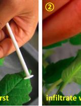
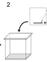
Quantification of Ethylene Production in Tomato Leaves Infected by Xanthomonas euvesicatoria
Preparation of Chloroplast Lipid Membrane and Lipid-protein Interaction Assay
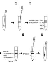
Measurement of PI4P Levels in Intact Chloroplasts Isolated from Arabidopsis thaliana
Stem Cell

Transfection of Embryoid Bodies with miRNA Precursors to Induce Cardiac Differentiation
