Past Issue in 2016

Volume: 6, Issue: 8

left

Dec

20

Dec

5

Nov

20

Nov

5

Oct

20

Oct

5

Sep

20

Sep

5

Aug

20

Aug

5

Jul

20

Jul

5

Jun

20

Jun

5

May

20

May

5

Apr

20

Apr

5

Mar

20

Mar

5

Feb

20

Feb

5

Jan

20

Jan

5

right

Biochemistry

HPLC Analysis of Secreted Organic Acids

HPLC Analysis of Secreted Organic Acids

Fiona K. Davies Fiona K. Davies
SD Sarah D’Adamo
MP Matthew C. Posewitz
10847 Views
Apr 20, 2016
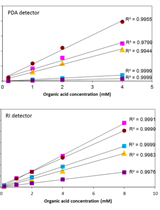
Under certain growth conditions some microorganisms secrete organic acids into the extracellular medium to relieve the accumulation of excess energy carriers, and/or to reduce toxic concentrations of organic acids. For example, a glycogen-deficient ∆glgC mutant of the cyanobacterium Synechococcus sp. PCC 7002 secretes pyruvate, acetate, α-ketoglutarate, α-ketoisocaproate and succinate (Davies et al., 2014; Jackson et al., 2015). Secretion of these organic acids functions as a putative energy-spilling mechanism in the absence of glycogen, the major carbon and reductant sink in this organism. Identification of secreted organic acids can facilitate the design of metabolic engineering strategies that funnel over-accumulating organic acids towards metabolic pathways that make a product of interest (such as a biofuel). Here, we describe a method for analyzing secreted organic acids in the extracellular media using high-performance liquid chromatography (HPLC). This method was developed for analysis of organic acids secreted by photosynthetic microbes (cyanobacteria and algae) into media, but could be used to analyze organic acids secreted by any microorganism cultivated in liquid medium.
Visualization of Intracellular Tyrosinase Activity in vitro

Visualization of Intracellular Tyrosinase Activity in vitro

RJ Riddhi Atul Jani
Sudeshna Nag Sudeshna Nag
Subba Rao Gangi Setty Subba Rao Gangi Setty
8825 Views
Apr 20, 2016
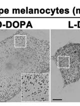
Melanocytes produce the melanin pigments in melanosomes and these organelles protect the skin against harmful ultraviolet rays. Tyrosinase is the key cuproenzyme which initiates the pigment synthesis using its substrate amino acid tyrosine or L-DOPA (L-3, 4-dihydroxyphenylalanine). Moreover, the activity of tyrosinase directly correlates to the cellular pigmentation. Defects in tyrosinase transport to melanosomes or mutations in the enzyme or reduced intracellular copper levels result in loss of tyrosinase activity in melanosomes, commonly observed in albinism. Here, we describe a method to detect the intracellular activity of tyrosinase in mouse melanocytes. This protocol will visualize the active tyrosinase present in the intracellular vesicles or organelles including melanosomes.

Microbiology

Measurement of Intracellular cAMP Levels Using the Cyclic Nucleotide XP Enzymatic Immunoassay Kit in Bacteria

Measurement of Intracellular cAMP Levels Using the Cyclic Nucleotide XP Enzymatic Immunoassay Kit in Bacteria

Sarah K Giles Sarah K Giles
Uwe H Stroeher Uwe H Stroeher
Melissa H Brown Melissa H Brown
11692 Views
Apr 20, 2016
Cyclic AMP (cAMP) is a ubiquitous secondary signaling molecule, commonly associated with many bacterial processes, including the regulation of virulence factors, such as biofilms, pellicles and motility (Wolfgang, 2003). The quantity of available cAMP is controlled by the interplay between the synthesis of adenosine triphosphate (ATP) to cAMP by adenylyl cyclases, and the degradation of cAMP by phosphodiesterase (McDonough et al., 2012). Adequate quantification of cAMP levels within a bacterial cell is an important step in identifying the impact that secondary signaling molecules play on the regulatory pathway within the cell. The principle of this method is to measure total cAMP levels within a bacterial cell, using crude bacterial whole cell lysate. The Cyclic AMP XPTM Assay kit used in this protocol was originally designed to be used for determining the level of cAMP in eukaryotic cells, however, the antibodies used in coating the wells will react with cAMP from any species and thus can be used for determining levels in bacterial cells. The measurement of cAMP in prokaryotic cells described here is a simple and cost effective method of producing quantifiable results.
Identification and Characterization of Bacterial Chemoreceptors Using Quantitative Capillary and Gradient Plate Chemotaxis Assays

Identification and Characterization of Bacterial Chemoreceptors Using Quantitative Capillary and Gradient Plate Chemotaxis Assays

JR Jose Antonio Reyes-Darias
Vanina García Vanina García
Miriam Rico-Jiménez Miriam Rico-Jiménez
Andrés Corral-Lugo Andrés Corral-Lugo
Tino Krell Tino Krell
11138 Views
Apr 20, 2016
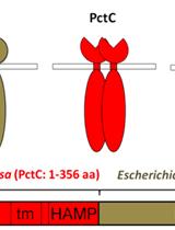
Bacterial chemotaxis is a motility-based response that biases cell movement toward beneficial molecules, called attractants, and away from harmful molecules, also known as repellents. Since the species of the genus Pseudomonas are characterized by a metabolic versatility, these bacteria have developed chemotactic behaviors towards a wide range of different compounds. The specificity of a chemotactic response is determined by the chemoreceptor, which is at the beginning of the signaling cascade and which receives the signal input. The basic elements of a typical chemoreceptor are the periplasmic ligand binding domain (LBD), responsible for sensing environmental stimuli, and the cytosolic methyl-accepting (MA) domain, that interacts with other components of the cellular signaling cascade. Escherichia coli (E. coli), the traditional model in chemotaxis research, has 5 well-characterized chemoreceptors. However, genome sequence analyses have revealed that many other bacteria possess many more chemoreceptors, some of which with partially overlapping signal profiles. This high number of chemoreceptors complicates their study by the analysis of single chemoreceptor mutants. We have pursued an alternative strategy for chemoreceptor characterization which corresponds to the generation of chimeric receptors composed of the LBD of the chemoreceptor under investigation and the MA domain of an E. coli receptor (Tar). The chimer is then introduced into a chemoreceptor free mutant of E. coli and the chemotaxis of the resulting strain is entirely due to the action of this chimeric receptor. In this publication we describe the use of quantitative capillary and gradient plate assays to study Pseudomonas chemotaxis as well as E. coli strains harboring chimeric receptors.
ACE-score-based Analysis of Temporal miRNA Targetomes  During Human Cytomegalovirus Infection Using AGO-CLIP-seq

ACE-score-based Analysis of Temporal miRNA Targetomes During Human Cytomegalovirus Infection Using AGO-CLIP-seq

SK Sungchul Kim
Kwangseog Ahn Kwangseog Ahn
10903 Views
Apr 20, 2016
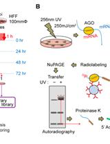
Although temporal regulation of gene expression during the course of infection is known to be critical for determining the outcome of host-virus interactions, systematic temporal analysis of the miRNA targetomes during productive viral infection has been technically challenging due to the large range of miRNA-mRNA cross-talks at the host-virus interface. High-confidence quantifying models of the suppression efficacy in targeting sites by integrating bioinformatics with Argonaute-crosslinking and immunoprecipitation followed by high-throughput sequencing (AGO-CLIP-seq) (Chi et al., 2009) data have been poorly developed. To accurately identify miRNA target sites and calculate the targeting efficacy of miRNA-target interactions, we developed a new bioinformatic quantitation method, AGO-CLIP-seq enrichment (ACE)-scoring algorithm (Kim et al., 2015). Inclusion of the uninfected control in our AGO-CLIP-seq analysis can significantly improve the accuracy of authentic target site identification for viral or human miRNAs and extract physiologically significant changes during productive human cytomegalovirus (HCMV) infection using our ACE-scoring method. Thus, we suggest that our new ACE-scoring-based methodology can be applied to various miRNA targetome studies, which will be performed in other kinds of temporal contexts, such as developmental stages, immune stimulation by cytokines or pathogens, and lytic infection by other viruses.

Neuroscience

Phagocytosis Assay of Microglia for Dead Neurons in Primary Rat Brain Cell Cultures

Phagocytosis Assay of Microglia for Dead Neurons in Primary Rat Brain Cell Cultures

Xiurong Zhao Xiurong Zhao
LZ Liyan Zhang
ST Shun-Ming Ting
JA Jaroslaw Aronowski
10471 Views
Apr 20, 2016
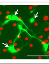
Clearance of dead brain tissue including the dead neurons through phagocytosis is an endogenous function of microglia in the brain, which is critical for inflammation resolution after ischemic stroke or head trauma. By regulating the function or polarization status of microglia, we may control their phagocytosis efficacy and therefore the cleanup process for the dead brain tissue. We cultured rat cortical neurons and microglia from the same litter of embryos. The cultured neurons are subjected to irradiation for inducing neuronal apoptosis. After labeling with propidium iodide (PI), the dead neurons (DNs) are exposed to the cultured microglia for phagocytosis assay. By counting the number of DNs in each microglia, we calculate the phagocytosis index to quantify the phagocytosis efficacy of microglia toward DNs. The protocol is divided into 4 sections: A) culturing rat cortical neurons from pre-natal rat embryos, B) preparing dead neurons as phagocytosis target, C) culturing rat brain microglia, D) quantifying phagocytosis index of microglia toward the dead neurons.
Primary Neuron-glia Culture from Rat Cortex as a Model to Study Neuroinflammation in CNS Injuries or Diseases

Primary Neuron-glia Culture from Rat Cortex as a Model to Study Neuroinflammation in CNS Injuries or Diseases

Ya-Ni Huang Ya-Ni Huang
Jia-Yi Wang Jia-Yi Wang
10450 Views
Apr 20, 2016
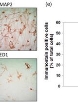
Primary neuron-glia cultures are commonly used in vitro model for neurobiological studies. Here, we provide a protocol for the isolation and culture of neuron-glial cells from cortical tissues of 1-day-old neonatal Sprague-Dawley pups. The procedure makes available an easier way to obtain the neuron and glia. In this culture system, neuron-glia cultures consisted of approximately 37% neurons, 51% astrocytes, 7% microglia, and a small percentage (<5%) of other cells after fourteen days in vitro. Primary neuron-glia cultures is a simplified in vitro model for studies focusing on interactions between neurons and glia cells. Activated glial cells, mainly astrocytes and microglia, are histopathological hallmarks of acute injury of the central nervous system (CNS) or chronic neurologic diseases (Hirsch and Hunot, 2009; Lee et al., 2009; Minghetti, 2005). Inflammatory mediators (e.g., nitric oxide, reactive oxygen species, proinflammatory cytokines, and chemokines) released by activated glia can directly or indirectly cause neuronal damage or neurodegeneration. Neuroinflammation is a common mechanism of various neurological diseases leading to neurodegeneration. The advantages of neuron-glia cultures are that: (1) Cultured cells can bypass complicated physiological interactions (such as leukocyte infiltration, blood-brain barrier, reflex or other systemic regulation) in vivo to allow direct observation of neuroinflammation caused by various CNS insults (hypoxia, ischemia, trauma. infection, neurotoxins, chronic stress or diseases); (2) Unlike cell lines that are mostly derived from tumor cells, primary cultured neuron-glia system is closer to the cell population ratio in vivo and can mimic the in situ microenvironment; and (3) Cultures can be prepared from various brain regions (e.g., cortex, hippocampus, mesencephalon…etc.) and allow an opportunity to examine the regional difference in the susceptibility to neurodegeneration following neuroinflammation caused by various CNS insults (Kim et al., 2000). The following protocol is an example for primary rat cortical neuron-glia culture preparation (Huang et al., 2015; Huang et al., 2014; Huang et al., 2012; Huang et al., 2009).
Aβ Extraction from Murine Brain Homogenates

Aβ Extraction from Murine Brain Homogenates

BC Brad T. Casali
GL Gary E. Landreth
10149 Views
Apr 20, 2016
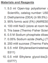
This protocol details beta-amyloid (Aβ) extraction from transgenic murine brain homogenates. Specifically, mechanical homogenization of brain tissue and sequential extraction of both soluble and insoluble proteins are detailed. DEA extracts soluble proteins, such as Aβ isoforms and APP. Formic acid enables extraction of insoluble protein aggregates, such as Aβ isoforms associated with plaques. This procedure produces soluble and insoluble extracts that are amenable to analysis of Aβ species via western blotting and/or enzyme-linked immunosorbent assays (ELISAs), and these results help assess amyloidogenic burden in animals.

Plant Science

Mating and Progeny Isolation in the Corn Smut Fungus Ustilago maydis

Mating and Progeny Isolation in the Corn Smut Fungus Ustilago maydis

MN Marina Nadal
JT Johanna E. Takach
DA David L. Andrews
SG Scott E. Gold
10340 Views
Apr 20, 2016
The corn smut pathogen, Ustilago maydis (U. maydis) (DC.) Corda, is a semi-obligate plant pathogenic fungus in the phylum Basidiomycota (Alexopoulos et al., 1996). The fungus can be easily cultured in its haploid yeast phase on common laboratory media. However, to complete its sexual cycle U. maydis strictly requires its specific plant host, maize (Zea mays). The fungus is an interesting and important model organism for the study of the interactions of fungal biotrophic pathogens with plants. In this protocol, we describe the process of plant inoculation, teliospore recovery, germination, progeny isolation and initial mating type analysis. The primary purpose of this protocol is to identify individual progeny strains of U. maydis that can be used for downstream genetic analyses. Generation of targeted mutants to study various processes is a common approach with this and many plant pathogenic fungi. The ability to generate combinations of mutations is facilitated by sexual crossing without the need for additional selectable markers.
Measuring the Interactions between Peroxisomes and Chloroplasts by in situ Laser Analysis

Measuring the Interactions between Peroxisomes and Chloroplasts by in situ Laser Analysis

Kazusato Oikawa Kazusato Oikawa
SM Shoji Mano
Kenji Yamada Kenji Yamada
YH Yoichiro Hosokawa
Mikio Nishimura Mikio Nishimura
7306 Views
Apr 20, 2016
Quantitative analysis has been necessary for deeply understanding characteristic of organelles function. This is the detailed protocol for the quantification of the physical interaction between peroxisomes and chloroplasts taken by laser scanning microscopy described by Oikawa et al. (2015). To clarify the morphological interactions between both organelles, we measured the contact length between two organelles (interaction length) in the fluorescent microscope image by using image analysis software ImageJ. The result clearly revealed that the contact length in light condition is much longer than that in dark condition. In addition, the force of the morphological interaction was quantified utilizing intersection technology of femtosecond laser and atomic force microscope (AFM). When an intense femtosecond laser is focused near the interface of two organelles, the adhesion is broken by a force due to the laser. The adhesion strength in light and dark conditions was estimated from the force calibrated by AFM. The detailed procedure is described in Bio-protocol as another protocol entitled “Quantification of the adhesion strength between peroxisomes and chloroplasts by femtosecond laser technology” (Hosokawa et al., 2016). These methods can be applied to other physical interaction between different types of organelles such as nuclei, mitochondria, Golgi, and chloroplasts.