
- Submit a Protocol
- Receive Our Alerts
- EN

- Protocols

- Articles and Issues
- For Authors
- About
- Become a Reviewer

Past Issue in 2016
Volume: 6, Issue: 13
Biochemistry
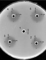
An Improved and Simplified Radial Gel Diffusion Assay for the Detection of Xylanase Activity


Cancer Biology
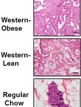
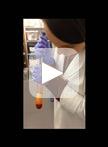
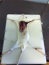
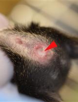
High Fat Diet-induced Breast Cancer Model in Rat

Cell Biology
Isolation and Primary Culture of Adult Mouse Cardiac Fibroblasts

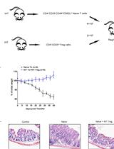
T Cell Transfer Model of Colitis

Subchromoplast Fractionation Protocol for Different Solanaceae Fruit Species


Developmental Biology
Aorta Ring Assay


Immunology
Mouse Embryonic Fibroblast Cell Culture and Stimulation
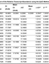
Measurement of mRNA Decay in Mouse Embryonic Fibroblasts
Molecular Biology
Estimation of the Chromosomal Copy Number in Synechococcus elongatus PCC 7942


Neuroscience
Locomotion Activity Measurement in an Open Field for Mice


Denervation of Mouse Lower Hind Limb by Sciatic and Femoral Nerve Transection

Plant Science
Indirect Immunofluorescence Assay in Chlamydomonas reinhardtii

Chromosome Dosage Analysis in Plants Using Whole Genome Sequencing


Identification of Natural Hybrids by SSR Markers in Mussaenda


Stem Cell
Retinal Differentiation of Mouse Embryonic Stem Cells
