
- Submit a Protocol
- Receive Our Alerts
- EN

- Protocols

- Articles and Issues
- For Authors
- About
- Become a Reviewer

Past Issue in 2016
Volume: 6, Issue: 22
Biochemistry
Ionization Properties of Phospholipids Determined by Zeta Potential Measurements


Determining Efficiency and Selectivity of Lipid Extraction by Perturbing Agents from Model Membranes


![Determination of Rate of [3H-methyl]-choline Incorporation into Cellular Lipids and Non-lipid Metabolites](https://en-cdn.bio-protocol.org/imageup/arcimg/20161108094711249.jpg?t=1758534059)
Determination of Rate of [3H-methyl]-choline Incorporation into Cellular Lipids and Non-lipid Metabolites
Cancer Biology

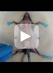
Establishment of Patient-Derived Xenografts in Mice


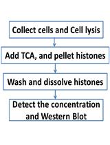
Acid Extraction of Total Histone from Colon Cancer HCT116 Cells
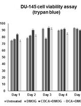
Determining the Influence of Small Molecules on Hypoxic Prostate Cancer Cell (DU-145) Viability Using Automated Cell Counting and a Cell Harvesting Protocol

Cell Biology
Quantitative Measurements of HIV-1 and Dextran Capture by Human Monocyte-derived Dendritic Cells (MDDCs)

Immunology
Isolation of Highly Pure Primary Mouse Alveolar Epithelial Type II Cells by Flow Cytometric Cell Sorting


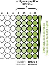
Evaluation of Cross-presentation in Bone Marrow-derived Dendritic Cells in vitro and Splenic Dendritic Cells ex vivo Using Antigen-coated Beads


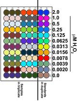
Determination of H2O2 Generation by pHPA Assay
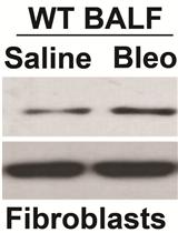
Assay to Evaluate BAL Fluid Regulation of Fibroblast α-SMA Expression
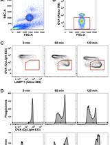
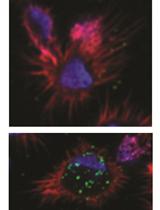
Analysis of Phagosomal Antigen Degradation by Flow Organellocytometry

Microbiology
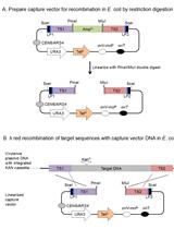
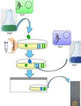
Transfer of Large Contiguous DNA Fragments onto a Low Copy Plasmid or into the Bacterial Chromosome
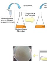
Transformation of Thermus Species by Natural Competence
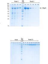
Heterologous Expression and Purification of the Magnesium Transporter A (MgtA) in Escherichia coli
Cell-to-cell DNA Transfer among Thermus Species
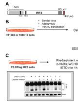
Biochemical Analysis of Caspase-8-dependent Proteolysis of IRF3 in Virus-infected Cells

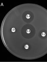
Halo Assay for Toxic Peptides and Other Compounds in Microorganisms


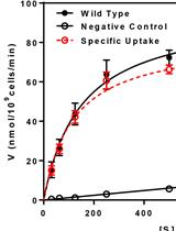
Uptake Assay for Radiolabeled Peptides in Yeast
Neuroscience
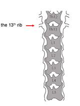
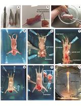
Microinjection of Virus into Lumbar Enlargement of Spinal Dorsal Horn in Mice
In vitro Brainstem-spinal Cord Preparation from Newborn Rat


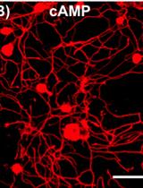
An in vitro Model of Neuron-macrophage Interaction to Generate Macrophages with Neurite Outgrowth Properties
Plant Science
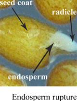
Arabidopsis Seed Germination Assay with Gibberellic Acid
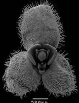
Documentation of Floral Secretory Glands in Pleurothallidinae (Orchidaceae) Using Scanning Electron Microscopy (SEM)
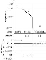
Cryopreservation Protocol for Chlamydomonas reinhardtii

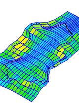
Visualising Differential Growth of Arabidopsis Epidermal Pavement Cells Using Thin Plate Spline Analysis


Stem Cell
Isolation and Primary Culture of Various Cell Types from Whole Human Endometrial Biopsies

Isolation and Culture of Human Adipose-derived Stem Cells from Subcutaneous and Visceral White Adipose Tissue Compartments


