
- Submit a Protocol
- Receive Our Alerts
- EN

- Protocols

- Articles and Issues
- For Authors
- About
- Become a Reviewer

Past Issue in 2017
Volume: 7, Issue: 2
Biochemistry
Assay to Access Anthelmintic Activity of Small Molecule Drugs Using Caenohabidtis elegans as a Model


Cancer Biology
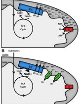
Assessment of Cellular Redox State Using NAD(P)H Fluorescence Intensity and Lifetime


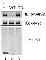
In vitro Dephosphorylation Assay of c-Myc

Developmental Biology
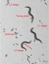
P-body and Stress Granule Quantification in Caenorhabditis elegans


Transplantation of Mesenchymal Cells Including the Blastema in Regenerating Zebrafish Fin
Immunology
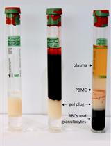
Isolation of PBMCs Using Vacutainer® Cellular Preparation Tubes (CPTTM)

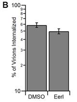
Virus Binding and Internalization Assay for Adeno-associated Virus

Microbiology
Aggregation Prevention Assay for Chaperone Activity of Proteins Using Spectroflurometry


Quantification of Triphenyl-2H-tetrazoliumchloride Reduction Activity in Bacterial Cells
Protein Expression Protocol for an Adenylate Cyclase Anchored by a Vibrio Quorum Sensing Receptor
Neuroscience
An Acute Mouse Spinal Cord Slice Preparation for Studying Glial Activation ex vivo


Plant Science
Nitrate Assay for Plant Tissues


Pseudomonas syringae Flood-inoculation Method in Arabidopsis


Two-electrode Voltage-clamp Recordings in Xenopus laevis Oocytes:Reconstitution of Abscisic Acid Activation of SLAC1 Anion Channel via PYL9 ABA Receptor


Bioassay of Xanthomonas albilineans Attachment on Sugarcane Leaves


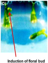
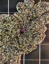
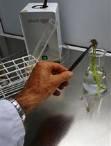
In vitro Floral Induction of Cuscuta reflexa
