
- Submit a Protocol
- Receive Our Alerts
- EN

- Protocols

- Articles and Issues
- For Authors
- About
- Become a Reviewer

Past Issue in 2017
Volume: 7, Issue: 4
Cancer Biology
Protocol for Murine/Mouse Platelets Isolation and Their Reintroduction in vivo


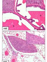
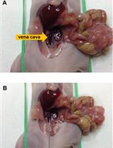
A Murine Orthotopic Allograft to Model Prostate Cancer Growth and Metastasis
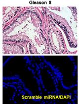
In situ Hybridization (ISH) and Quantum Dots (QD) of miRNAs


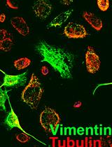
Analysis of Cancer Stromal Reaction Using an O-ring Co-culture Assay
miRNA Characterization from the Extracellular Vesicles
Cell Biology
Quantitative Analysis of Exosome Secretion Rates of Single Cells
Microbiology
Expression and Purification of Cyanobacterial Circadian Clock Protein KaiC and Determination of Its Auto-phosphatase Activity


Molecular Biology
Production of Guide RNAs in vitro and in vivo for CRISPR Using Ribozymes and RNA Polymerase II Promoters
Plant Science
Automatic Quantification of the Number of Intracellular Compartments in Arabidopsis thaliana Root Cells


Endophytic Microbial Community DNA Extraction from the Plant Phyllosphere
Penetration Assays, Fungal Recovery and Pathogenicity Assays for Verticillium dahliae
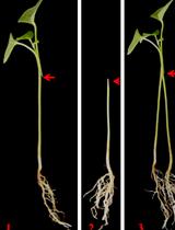
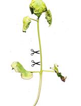
Establishment of New Split-root System by Grafting

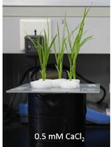
Rice Root Organic Acid Efflux Measurement by Using Ion Chromatography


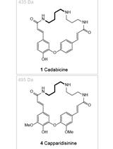
Chromatographic Separation of the Codonocarpine Type Alkaloids from the Root Bark of Capparis decidua


Stem Cell
Adhesion Assay for Murine Bone Marrow Hematopoietic Stem Cells
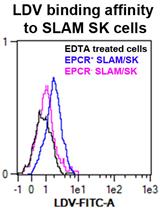
VLA-4 Affinity Assay for Murine Bone Marrow-derived Hematopoietic Stem Cells