
- Submit a Protocol
- Receive Our Alerts
- EN

- Protocols

- Articles and Issues
- For Authors
- About
- Become a Reviewer

Past Issue in 2017
Volume: 7, Issue: 5
Cancer Biology
Liposome Flotation Assays for Phosphoinositide-protein Interaction
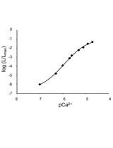
Ca2+ Measurements in Mammalian Cells with Aequorin-based Probes
Developmental Biology
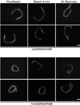
Protein Synthesis Rate Assessment by Fluorescence Recovery after Photobleaching (FRAP)

Microbiology
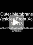
Isolation of Outer Membrane Vesicles from Phytopathogenic Xanthomonas campestris pv. campestris
Transient Transfection-based Fusion Assay for Viral Proteins

Determination of Elemental Concentrations in Lichens Using ICP-AES/MS


Next-generation Sequencing of the DNA Virome from Fecal Samples


Polyethylene Glycol-mediated Transformation of Drechmeria coniospora
An HPLC-based Method to Quantify Coronatine Production by Bacteria


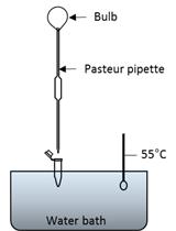
Chitin Extraction and Content Measurement in Magnaporthe oryzae

Neuroscience
Extracellular Axon Stimulation


Axonal Conduction Velocity Measurement

Olfactory Habituation-dishabituation Test (Mouse)

Olfactory Avoidance Test (Mouse)
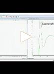
Thinned-skulled Cranial Window Preparation (Mice)
Plant Science
Acetyl Bromide Soluble Lignin (ABSL) Assay for Total Lignin Quantification from Plant Biomass

In Gel Kinase Assay


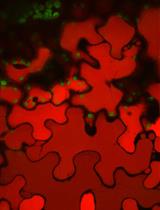
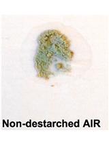
Laser Scanning Confocal Microcopy for Arabidopsis Epidermal, Mesophyll, and Vascular Parenchyma Cells
Surface Inoculation and Quantification of Pseudomonas syringae Population in the Arabidopsis Leaf Apoplast
Multiplexed GuideRNA-expression to Efficiently Mutagenize Multiple Loci in Arabidopsis by CRISPR-Cas9
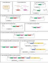
Knock-in Blunt Ligation Utilizing CRISPR/Cas9

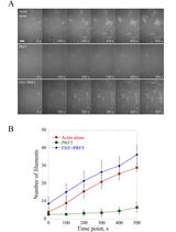
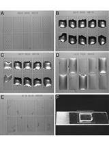
Direct Visualization and Quantification of the Actin Nucleation and Elongation Events in vitro by TIRF Microscopy

Stem Cell
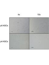
Isolation and Primary Culture of Adult Human Adipose-derived Stromal/Stem Cells


Reprogram Murine Epiblast Stem Cells by Epigenetic Inhibitors