
- Submit a Protocol
- Receive Our Alerts
- EN

- Protocols

- Articles and Issues
- For Authors
- About
- Become a Reviewer

Past Issue in 2013
Volume: 3, Issue: 16
Biochemistry
Protein Flotation Assay to Isolate Lipids Rafts from Soft Tissue or Cells


PTEN-lipid Binding Assay
Cancer Biology
Quantitative Methylation Specific PCR (qMSP)
Natural Killer Cell Transfer Assay


Cell Biology
Cell Surface Protein Biotinylation and Analysis


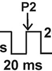
Sodium Current Measurements in HEK293 Cells
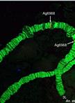
Fluorescence in situ Hybridization to the Polytene Chromosomes of Anopheles Mosquitoes
Immunology
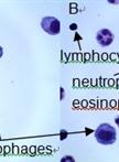
Bronchoalveolar Lavage and Lung Tissue Digestion

Ex vivo Natural Killer Cell Cytotoxicity Assay
Microbiology
Zymogram Assay for the Detection of Peptidoglycan Hydrolases in Streptococcus mutans

Drug Sensitivity Assay of Xanthomonas citri subsp. citri Using REMA Plate Method

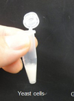
![Metabolic Labeling of Yeast Sphingolipids with Radioactive D-erythro-[4,5-3H]dihydrosphingosine](https://en-cdn.bio-protocol.org/imageup/arcimg/20130820012439267.jpg?t=1758534032)
Metabolic Labeling of Yeast Sphingolipids with Radioactive D-erythro-[4,5-3H]dihydrosphingosine
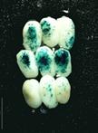
Bioassay to Screen Pathogenesis-associated Genes in Alternaria brassicicola
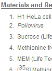
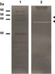
Isolation of Radiolabeled Poliovirus Particles from H1 HeLa Cells
Molecular Biology
Ultra-low Background DNA Cloning System

Neuroscience
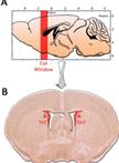
Retrograde and Anterograde Tracing of Neural Projections


Primary Culture of SVZ-derived Progenitors Grown as Neurospheres
Plant Science
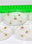
Measuring Germination Percentage in Wheat
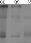
Reverse Zymmogram Analysis for the Detection of Protease Inhibitor Activity

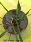
Rapid Induction of Water Stress in Wheat
Maize Embryo Transient Transformation by Particle Bombardment
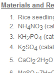
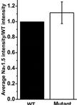
Determination of Nitrate Uptake and Accumulation Using 15N in Rice Seedlings


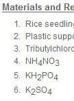
Measurement of Net NO3- Flux in Rice Plants with the SIET System