
- Submit a Protocol
- Receive Our Alerts
- EN

- Protocols

- Articles and Issues
- For Authors
- About
- Become a Reviewer

Past Issue in 2013
Volume: 3, Issue: 18
Biochemistry
Immunoprecipitation of ROR1
Cancer Biology
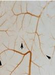
In vivo Chick Chorioallantoic Membrane (CAM) Angiogenesis Assays

Neutral Comet Assay


Isolation of Mouse Embryo Fibroblasts
Homologous Recombination Assay
End-synapsis Assay
Cell Biology
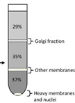
Enrichment of Golgi membranes from HeLa cells by sucrose gradient ultracentrifugation
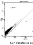
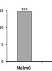
ROR1 Flow Cytometry
Immunology
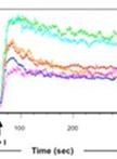
Flow Cytometric Analysis of Calcium Influx Assay in T cells

Microbiology
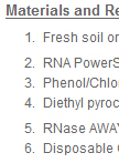
Isolating RNA from the Soil
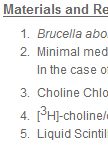
Choline Uptake Assay in Bacterial Cells

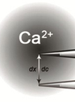
Measurement of Extracellular Ca2+ Influx and Intracellular H+ Efflux in Response to Glycerol and PEG6000 Treatments


Molecular Biology
Chromatin Immunoprecipitation (ChIP), Streptavidin and ATP-agarose Mediated Pull-down Analyses

Neuroscience
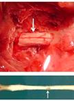
Rat Model of Chronic Midthoracic Lateral Hemisection

Plant Science
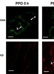
Fluorescence Measurement of Postharvest Physiological Deterioration (PPD) in Cassava Storage Roots


Analysis of Flower Cuticular Waxes and Cutin Monomers

Stem Cell
iPS Cell Induction from Human Non-T, B cells from Peripheral Blood