
- Submit a Protocol
- Receive Our Alerts
- EN

- Protocols

- Articles and Issues
- For Authors
- About
- Become a Reviewer

Past Issue in 2013
Volume: 3, Issue: 22
Cell Biology
Harvest and Culture of Mouse Peritoneal Macrophages

Immunology
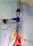
Gastric Aspiration Models

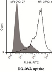
Assessment of Human Dendritic Cell Antigen Uptake by Flow Cytometry
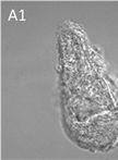
Immunocytochemical Detection of Recombinant Biomphalysin on Schistosoma mansoni Sporocysts

Microbiology
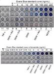
Assay to Evaluate Vascular Permeability Induction in Mice


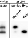
Separation and Detection of Phosphorylated and Nonphosphorylated BvgA, a Bordetella pertussis Response Regulator, in vivo and in vitro
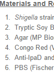
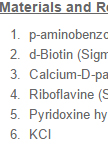
Shigella IpaD and IpaB Surface Localizations

Transport Assays in Aspergillus nidulans


Neuroscience
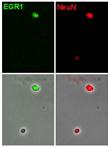
Cell Cycle Analysis in the Vertebrate Brain Using Immunolabeled Fresh Cell Nuclei


Plant Science

Analysis of RNA-protein Interactions Using Electrophoretic Mobility Shift Assay (Gel Shift Assay)

Bimolecular Fluorescence Complementation (BIFC) Protocol for Rice Protoplast Transformation

Shikimate Hydroxycinnamoyl Transferase (HCT) Activity Assays in Populus nigra

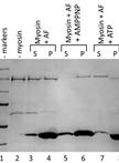
Binding Assay of Cytosolic Proteins to the Cytoskeleton


Mapping and Analysis of Illumina Reads for Transcriptome of Medicago Truncatula During the Early Organogenesis of the Nodule
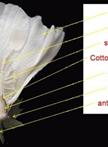
Cotton Ovules Culture and Analysis
