Improve Research Reproducibility A Bio-protocol resource

- Submit a Protocol
- Receive Our Alerts
- EN

- Protocols

- Articles and Issues
- For Authors
- About
- Become a Reviewer

Past Issue in 2014
Volume: 4, Issue: 2
Biochemistry
A Protocol for Measurement of Intracellular pH


Intracellular pH (pHi) is an important physiological determinant of enzyme activity and cellular function (Kurkdjian and Guern, 1989). All proteins depend on a tightly regulated pH to maintain their structure and function. Protonation–deprotonation events can dictate the charge of biological surfaces and are integral steps in many metabolic reactions (Casey et al., 2010). Moreover, the proton gradient across the mitochondrial membrane is used to generate cellular energy and support other mitochondrial processes. As a result, cells have developed multiple mechanisms to maintain a narrow range of pHi in response to extra- and intracellular fluctuations in pH (Orij et al., 2012). Here, we describe a protocol for pHi measurement in live cells that uses fluorescent microscopy and the pH sensitive dye 2’,7’-Bis-(2-Carboxyethyl)-5-(and-6-)-Carboxyfluorescein Acetoxymethyl Ester (BCECF-AM). This method was recently used to determine the effects of intracellular pH changes on global histone acetylation levels (McBrian et al., 2013).
Cell Biology
Preparation of Bacillus subtilis Cell Lysates and Membranes
A common feature of every eukaryotic and prokaryotic cell is that they exhibit a plasma membrane. In Bacillus subtilis (B. subtilis) roughly 25% of all proteins are putative trans- or membrane associated proteins. Here we describe a relatively simple method to separate and prepare membrane and cytosolic proteins by ultra-centrifugation.
Immunology
ADCC Assay Protocol
Antibody-dependent cell-mediated cytotoxicity (ADCC) bridges innate and adaptive immunity, and it involves both humoral and cellular immune responses. ADCC has been found to be a main route of immune protection against viral infections and cancers in vivo. Here we developed a flow cytometry based protocol for ADCC assay using human peripheral blood mononuclear cells (PBMCs) as effector cells. Using this protocol, we determined the ADCC activity of convalescent plasma IgGs from six H1N1-infected human subjects in China, and identified two dominant ADCC epitopes, designated E1 [amino acid (AA) 92-117] and E2 (AA 124-159), on haemagglutinin of pandemic H1N1 influenza virus by epitope mapping of the convalescent plasma IgGs with different levels of ADCC activity. Our study may aid in designing immunogens that can elicit antibodies with high ADCC activity. Vaccine immunogens designed to include the structural determinants of potent broadly neutralizing antibodies and ADCC epitopes may confer a comprehensive immune protection against viral infections.
Microbiology
Isolation of the Secretome from Bacillus subtilis
Bacteria are commonly known to secret proteins in large amounts into the surrounding environment in high concentrations via various pathways. These proteins can be involved in numerous processes like cell-cell communication, exopolymer formation but also metabolic active enzymes are secreted that are interesting for industrial production of proteins. One of the most regularly used organisms for industrial protein production is the Gram-positive bacterium Bacillus subtilis (B. subtilis). Here we describe a protocol that can be used to quantitatively and qualitatively analyze secreted proteins from B. subtilis.
Biochemical Assays for MTase Activity

Methyltransferase (MTase) transfers a methyl group (-CH3) from the donor S-adenosyl-L-methionine (AdoMet or SAM) to biologically active molecules such as hormones, neurotransmitters, lipids, proteins and nucleic acids. The addition of a methyl group causes a change in the physicochemical properties of the molecules. The mRNA cap structure is essential for cell and virus. Guanine-N7-methyltransferase (N7-MTase) methylates the GpppN cap at the N7 position of guanine, resulting in cap-0 structure (m7GpppN), and Ribose 2'-O-MTase further methylates the first nucleotide of higher eukaryotic cellular and viral mRNAs at the ribose 2'-OH position to form cap-1 (m7GpppNm) structures. Here, we describe a biochemical assay to detect the activities of mRNA capping MTases.
Neuroscience
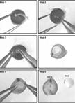
Retinal Explant Culture

A particularly powerful culture method for the retina is the explant assay, which consists in culturing a small piece of retina on an organotypic filter. Retinal explants can be prepared any time between embryonic day 13 (E13) and postnatal day 4 (P4). Although retinal ganglion cells tend to degenerate shortly after they are generated in explants, and photoreceptor cells do not grow extended outer segments, the explants will develop very similarly to a retina in vivo and generate all the different retinal cell types that will migrate to the appropriate layer. The retinal explant culture assay is particularly useful in cases where a mouse mutant is embryonic lethal and its retinal development cannot be studied in vivo. Because retinal explants can be prepared from embryonic animals and electroporated or infected with viral vectors, it is also a useful approach for the study of gene function at embryonic stages. Here, we present a retinal explant culture method that we have used extensively in various publications (Kechad et al., 2012; Cayouette et al., 2003; Cayouette and Raff, 2003; Elliott et al., 2008).
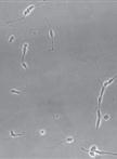
Dissociated Retinal Cell Culture
The retina is a relatively simple and accessible part of the central nervous system, making it a powerful model to study cell fate specification mechanisms. Multipotent retinal progenitor cells (RPCs) give rise to seven major classes of retinal cell types. Mechanisms regulating cell fate choice in the retina depend on both cell intrinsic and environmental factors, but their relative contribution to specific cell fate decisions remains unclear. Dissociated retinal cell cultures provide a great assay to study this problem. RPCs are cultured in serum-free and extract-free medium, providing the investigator with a control over the environment to address questions related to the effects of a particular molecule on the development of retinal neurons. In addition, dissociated cell cultures can be used to study the importance of cell intrinsic mechanisms by isolating RPCs from their normal environment (Cayouette et al., 2003; Jensen and Raff, 1997). The method described below is suitable for the clonal-density culture of RPCs. In such cultures, RPCs are isolated from each other and from the postmitotic neurons. They divide and differentiate into different retinal cell types to form small colonies, or “clones”. In a recent study, we found that these clones are indistinguishable from the clones that develop in situ in the retina, both in terms of cell number and cell type composition, suggesting that intracellular mechanisms play a key role in retinal development (Cayouette et al., 2003).
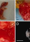
Neuron Culture from Mouse Superior Cervical Ganglion
The rodent superior cervical ganglion (SCG) is a useful and readily accessible source of neurons for studying the mechanisms of sympathetic nervous system (SNS) development and growth in vitro. The sympathetic nervous system (SNS) of early postnatal animals undergoes a great deal of remodeling and development; thus, neurons taken from mice at this age are primed to re-grow and establish synaptic connections after in situ removal. The stereotypic location and size of the SCG make it ideal for rapid isolation and dissociation. The protocol described here details the requirements for the dissection, culture and differentiation of SCG neurons. The protocol is suitable for culturing neurons from late embryonic gestation to approximately postnatal day 3. The culture technique discussed below utilizes glass coverslips for the microscopic examination of fixed cells.
Proteasome Assay in Cell Lysates

The ubiquitin-proteasome system (UPS) mediates the majority of the proteolysis seen in the cytoplasm and nucleus of mammalian cells. As such it plays an important role in the regulation of a variety of physiological and pathophysiological processes including tumorigenesis, inflammation and cell death (Ciechanover, 2005; Kisselev and Goldberg, 2001). A number of recent studies have shown that proteasome activity is decreased in a variety of neurological disorders including Parkinson's disease, Alzheimer's disease, amyotrophic lateral sclerosis and stroke as well as during normal aging (Chung et al., 2001; Ciechanover and Brundin, 2003; Betarbet et al., 2005). This decrease in proteasome activity is thought to play a critical role in the accumulation of abnormal and oxidized proteins. Protein clearance by the UPS involves two sequential reactions. The first is the tagging of protein lysine residues with ubiquitin (Ub) and the second is the subsequent degradation of the tagged proteins by the proteasome. We herein describe an assay for the second of these two reactions (Valera et al., 2013). This assay uses fluorogenic substrates for each of the three activities of the proteasome: chymotrypsin-like activity, trypsin-like activity and caspase-like activity. Cleavage of the fluorophore from the substrate by the proteasome results in fluorescence that can be detected with a fluorescent plate reader.
Monocular Deprivation in Mice

Monocular deprivation is an experimental technique to study the ocular dominance plasticity during critical period (Hubel and Wiesel, 1963). Generally one eye of an animal is sutured during critical period, and the sutured eye is re-opened after either less than three days (short term) or more than three days (long term). Here we describe a detailed protocol for short-term and long-term monocular deprivation in mouse (Ma et al., 2013).
Plant Science
Monoclonal Antibody Purification (Nicotiana benthamiana Plants)


Plant-based expression systems provide an alternative biomanufacturing platform for recombinant proteins (Matoba et al., 2011). In particular, plant virus-based vectors can overexpress proteins within days in the leaf tissue of Nicotiana benthamiana (N. benthamiana). To overcome the issues of genetic instability and limited infectivity of recombinant viruses, Agrobacterium-mediated delivery of “deconstructed” virus vectors has become the mainstay for the production of large and/or multicomponent proteins, such as immunoglobulin (Ig)G monoclonal antibodies (mAbs). Here, we describe a method of producing human IgG mAbs in N. benthamiana using the tobamoviral replicon vector magnICON®. The vector can express up to a few hundred mg of a mAb per kg of leaf material in 7 days. A representative case for the broadly neutralizing anti-HIV and anti-influenza mAbs, VRC01 and CR6261 respectively, is shown (Hamorsky et al., 2013). Leaf tissue is homogenized and the extract is clarified by filtration and centrifugation. The mAb is purified by fast protein liquid chromatography (FPLC) using Protein A affinity and Phenyl HP hydrophobic interection resins.