Past Issue in 2014

Volume: 4, Issue: 8

left

Dec

20

Dec

5

Nov

20

Nov

5

Oct

20

Oct

5

Sep

20

Sep

5

Aug

20

Aug

5

Jul

20

Jul

5

Jun

20

Jun

5

May

20

May

5

Apr

20

Apr

5

Mar

20

Mar

5

Feb

20

Feb

5

Jan

20

Jan

5

right

Cancer Biology

In vivo Lineage-tracing Studies in a Cancer Stem Cell Population in Neuroblastoma

In vivo Lineage-tracing Studies in a Cancer Stem Cell Population in Neuroblastoma

DH Danielle M. Hsu
JS Jason M. Shohet
Eugene S.  Kim Eugene S. Kim
9264 Views
Apr 20, 2014
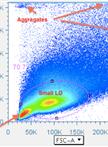
Tumors are comprised of heterogeneous subpopulations that may exhibit differing capacity for differentiation, self-renewal, and tumorigenicity. In vivo lineage-tracing studies are a powerful tool for defining the role of tumor subpopulations in tumor growth and as targets for therapeutic agents. This protocol describes using a neuroblastoma cancer cell line transduced with two different fluorescent proteins (GFP and tdTomato) to track the specific contributions of cells expressing the GCSF receptor (CD114+) or not (CD114-) on tumor growth in vivo.

Cell Biology

Purification and Fluorescent Labeling of Exosomes

Purification and Fluorescent Labeling of Exosomes

AN Asuka Nanbo
EK Eri Kawanishi
RY Ryuji Yoshida
HY Hironori Yoshiyama
19539 Views
Apr 20, 2014
Exosomes are small membrane vesicles of endocytic origin secreted into the extracellular environment from a variety of different cells, and are thought to play important roles in intercellular communications. Here, we provide a useful protocol to purify the exosomes released from cell lines using sucrose gradient centrifugation. In this protocol, we also applied a red-fluorescent lipophilic dye, DiI, which is incorporated in the outer membrane of exosomes. This fluorescently labeled exosomes allow us to visualize individual exosomes by a confocal laser scanning microscope.

Immunology

Quantification of T Cell Antigen-specific Memory Responses in Rhesus Macaques, Using Cytokine Flow Cytometry (CFC, also Known as ICS and ICCS): from Assay Set-up to Data Acquisition

Quantification of T Cell Antigen-specific Memory Responses in Rhesus Macaques, Using Cytokine Flow Cytometry (CFC, also Known as ICS and ICCS): from Assay Set-up to Data Acquisition

Andrew W. Sylwester Andrew W. Sylwester
SH Scott G. Hansen
LP Louis J. Picker
15763 Views
Apr 20, 2014
What was initially termed ‘CFC’ (Cytokine Flow Cytometry’) is now more commonly known as ‘ICS’ (Intra Cellular Staining), or less commonly as ‘ICCS’ (Intra Cellular Cytokine Staining). The key innovations were use of an effective permeant (allowing intracellular staining), and a reagent to disrupt secretion (trapping cytokines, thereby enabling accumulation of detectable intracellular signal). Because not all researchers who use the technique are interested in cytokines, the ‘ICS’ term has gained favor, though ‘CFC’ will be used here. CFC is a test of cell function, exposing lymphocytes to antigen in culture, then measuring any cytokine responses elicited. Test cultures are processed so as to stain cells with monoclonal antibodies tagged with fluorescent markers, and to chemically fix the cells and decontaminate the samples, using paraformaldehyde. CFC provides the powers of flow cytometry, which includes bulk sampling and multi-parametric cross-correlation, to the analysis of antigen-specific memory responses. A researcher using CFC is able to phenotypically characterize cells cultured with test antigen, and for phenotypic subsets (e.g. CD4+ or CD8+ T cells) determine the % frequency producing cytokine above background level.In contrast to ELISPOT and Luminex methods, CFC can correlate production of multiple cytokines from particular, phenotypically-characterized cells. The CFC assay is useful for detecting that an individual has had an antigen exposure (as in population screenings), or for following the emergence and persistence of antigen memories (as in studies of vaccination, infections, or pathogenesis). In addition to quantifying the % frequency of antigen-responding cells, mean fluorescence intensity can be used to assess how much of a cytokine is generated within responding cells.With the technological advance of flow cytometry, a current user of CFC often has access to 11 fluorescent channels (or even 18), making it possible to either highly-characterize the phenotypes of antigen-responding cells, or else simultaneously quantify the responses according to many cytokines or activation markers. Powerful software like FlowJo (TreeStar) and SPICE (NIAID) can be used to analyse the data, and to do sophisticated multivariate analysis of cytokine responses.The method described here is customized for cells from Rhesus macaque monkeys, and the extensive annotating notes represent a decade of accumulated technical experience. The same scheme is readily applicable to other mammalian cells (e.g. human or mouse), though the exact antibody clones will differ according to host system. The basic method described here incubates 1 x 10⁶ Lymphocytes in 1 ml tube culture with antigen and co-stimulatory antibodies in the presence of Brefeldin A, prior to staining and fixation.
Quantification of T Cell Antigen-specific Memory Responses in Rhesus Macaques, Using Cytokine Flow Cytometry (CFC, also Known as ICS and ICCS): Analysis of Flow Data

Quantification of T Cell Antigen-specific Memory Responses in Rhesus Macaques, Using Cytokine Flow Cytometry (CFC, also Known as ICS and ICCS): Analysis of Flow Data

Andrew W. Sylwester Andrew W. Sylwester
SH Scott G. Hansen
LP Louis J. Picker
15546 Views
Apr 20, 2014
What was initially termed ‘CFC’ (Cytokine Flow Cytometry) is now more commonly known as ‘ICS’ (Intra Cellular Staining), or less commonly as ‘ICCS’ (Intra Cellular Cytokine Staining). The key innovations were use of an effective permeant (allowing intracellular staining), and a reagent to disrupt secretion (trapping cytokines, thereby enabling accumulation of detectable intracellular signal). Because not all researchers who use the technique are interested in cytokines, the ‘ICS’ term has gained favor, though ‘CFC’ will be used here. CFC is a test of cell function, exposing lymphocytes to antigen in culture, then measuring any cytokine responses elicited. Test cultures are processed so as to stain cells with monoclonal antibodies tagged with fluorescent markers, and to chemically fix the cells and decontaminate the samples, using paraformaldehyde. CFC provides the powers of flow cytometry, which includes bulk sampling and multi-parametric cross-correlation, to the analysis of antigen-specific memory responses. A researcher using CFC is able to phenotypically characterize cells cultured with test antigen, and for phenotypic subsets (e.g. CD4+ or CD8+ T cells) determine the % frequency producing cytokine above background level.In contrast to ELISPOT and Luminex methods, CFC can correlate production of multiple cytokines from particular, phenotypically-characterized cells. The CFC assay is useful for detecting that an individual has had an antigen exposure (as in population screenings), or for following the emergence and persistence of antigen memories (as in studies of vaccination, infections, or pathogenesis). In addition to quantifying the % frequency of antigen-responding cells, mean fluorescence intensity can be used to assess how much of a cytokine is generated within responding cells.With the technological advance of flow cytometry, a current user of CFC often has access to 11 fluorescent channels (or even 18), making it possible to either highly-characterize the phenotypes of antigen-responding cells, or else simultaneously quantify the responses according to many cytokines or activation markers. Powerful software like FlowJo (TreeStar) and SPICE (NIAID) can be used to analyse the data, and to do sophisticated multivariate analysis of cytokine responses.The method described here is customized for cells from Rhesus macaque monkeys, and the extensive annotating notes represent a decade of accumulated technical experience. The same scheme is readily applicable to other mammalian cells (e.g. human or mouse), though the exact antibody clones will differ according to host system. The basic method described here incubates 1 x 10⁶ Lymphocytes in 1 ml tube culture with antigen and co-stimulatory antibodies in the presence of Brefeldin A, prior to staining and fixation.Note: This is the second part of a two-part procedure. Part one has the same initial title, but the subtitle “From assay set-up to data acquisition (Sylwester et al., 2014)”. The Abstract and Historical Background is the same for both documents.

Microbiology

Radioactive Pulse-Chase Analysis and Immunoprecipitation

Radioactive Pulse-Chase Analysis and Immunoprecipitation

Javier G.  Magadán Javier G. Magadán
12080 Views
Apr 20, 2014
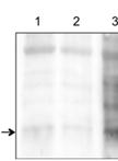
Labeling of newly-synthesized polypeptides with radioactive amino acids followed by immunoprecipitation allows quantitative analysis of the fate of a given protein in a time-dependent manner. This biochemical approach is usually used to study a variety of processes, such as protein folding, co-translational modifications, intracellular transport, and even its rate of degradation. Here, I describe step by step a simple technique to both label newly-synthesized influenza A virus (IAV) hemagglutinin (HA) with [35S]-methionine and then follow its maturation and transport through the secretory pathway by SDS-PAGE and fluorography (Magadan et al., 2013).
Assay for Adherence of Vibrio cholerae to Eukaryotic Cell Lines

Assay for Adherence of Vibrio cholerae to Eukaryotic Cell Lines

Amit K.  Dey Amit K. Dey
AB Abha Bhagat
RC Rukhsana Chowdhury
10598 Views
Apr 20, 2014
Vibrio cholerae (V. cholerae) colonizes the intestinal epithelium and secretes cholera toxin (CT), a potent enterotoxin that causes severe fluid loss characteristic of the disease cholera. V. cholerae is a non-invasive Gram-negative bacterium that adheres to intestinal cells as well as a variety of different cell types. A protocol for adherence of V. cholerae to various cell lines is described. The adhered bacteria can be used to examine expression of genes that are differentially expressed between adhered and unadhered bacteria or other purposes (Dey et al., 2013).
Purification and Crystallization of Chloromuconolactone Dehalogenase ClcF from Rhodococcus opacus 1CP

Purification and Crystallization of Chloromuconolactone Dehalogenase ClcF from Rhodococcus opacus 1CP

CR Christian Roth
JG Janosch Alexander D. Gröning
SK Stefan Rudolf Kaschabek
MS Michael Schlömann
NS Norbert Sträter
10493 Views
Apr 20, 2014
The protocol describes the generation of variants of chloromuconolactone dehalogenase from Rhodococcus opacus (R. opacus) 1CP. ClcF is a multimeric protein, which catalyses the dechlorination of 5-chloromuconolactone to cis-dienelactone in the 3-chlorocatecholic acid degradation pathway. The protocol describes the workflow for the purification and subsequent crystallization of the enzyme. The used workflow and the described techniques could be easily adapted to any other protein/enzyme intended to be crystallized by the potential user for subsequent structure determination. The protocol does not involve expensive specialized equipment which allows the use in standard laboratories not specially dedicated to macromolecular crystallography.
Zonal Sedimentation Analysis on Sucrose Gradients

Zonal Sedimentation Analysis on Sucrose Gradients

Javier G.  Magadán Javier G. Magadán
9262 Views
Apr 20, 2014
Zonal sedimentation analysis on sucrose gradients allows estimation of the molecular size of an individual protein or a protein complex by centrifugation at a constant speed under nondenaturing conditions. This method is particularly suitable for globular proteins like the influenza A virus (IAV) protein hemagglutinin (HA). Here, I describe step by step a protocol used to evaluate the oligomeric state of recombinant HA trimers (Magadan et al., 2013).
The BhbA Enzyme Assay

The BhbA Enzyme Assay

KC Kai Chen
Jiandong  Jiang Jiandong Jiang
8279 Views
Apr 20, 2014
Reductive dehalogenation has been found primarily in anaerobic communities and is originally thought to rarely occur in aerobes. A reductive dehalogenase (BhbA) was characterized from an aerobic strain of Comamonas sp. 7D-2, which was isolated from a bromoxynil octanoate-contaminated soil sample collected in Jiangsu, China. BhbA catalyzes the reductive dehalogenation of bromoxynil and its derivative 3,5-dibromo-4-hydroxybenzoate under aerobic conditions. BhbA is membrane-associated and found to have the key features of anaerobic respiratory reductive dehalogenases. This protocol describes the method for enzyme analysis of the aerobic reductive dehalogenase (BhbA) in the membrane fraction.

Plant Science

Histochemical Detection of Superoxide and H2O2 Accumulation in Brassica juncea Seedlings

Histochemical Detection of Superoxide and H2O2 Accumulation in Brassica juncea Seedlings

Deepak  Kumar Deepak Kumar
Mohd  Aslam  Yusuf Mohd Aslam Yusuf
Preeti  Singh Preeti Singh
Meryam  Sardar Meryam Sardar
Neera Bhalla Sarin Neera Bhalla Sarin
32094 Views
Apr 20, 2014
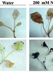
Plant cells continually produce reactive oxygen species (ROS) as a by-product of aerobic metabolism. Increased production of ROS occurs under unfavorable conditions imposed by various abiotic and biotic factors. Accumulation of ROS is damaging to various cellular components and macromolecules including plasma membrane, nucleic acids, and proteins and eventually leads to cell death. In this protocol, we describe the histochemical detection of hydrogen peroxide (H2O2) and superoxide (O2-) anion, two of the most important ROS, in Brassica juncea seedlings by using 3,3ʹ-Diaminobenzidine (DAB) and Nitrotetrazolium blue chloride (NBT) as the chromogenic substrate. DAB is oxidized by H2O2 in the presence of peroxidases and produces reddish brown precipitate. NBT reacts with O2- to form a dark blue insoluble formazan compound. The protocol can be used in other plant species and for different plant tissues.
Sandwich Enzyme-linked Immunosorbent Assay (ELISA) Analysis of Plant Cell Wall Glycan Connections

Sandwich Enzyme-linked Immunosorbent Assay (ELISA) Analysis of Plant Cell Wall Glycan Connections

Valérie   Cornuault Valérie Cornuault
JK J. Paul Knox
16424 Views
Apr 20, 2014
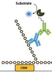
Sandwich ELISA is a highly sensitive method that can be used to determine if two epitopes are part of the same macromolecule or supramolecular complex. In the case of plant cell wall glycans, it can reveal the existence of inter-polymers linkages, leading to better understanding of overall cell wall architectures. This development of a conventional sandwich ELISA protocol uses a carbohydrate-binding module (CBM), a small protein domain found in some carbohydrate catalysing or activating enzymes, and rat monoclonal antibodies (mAbs) which can be combined in the same ELISA plate without risk of cross reaction; the secondary anti-rat HRP antibody being only able to bind to the rat mAb and not the CBM. This protocol was developed and modified in the Prof. J. Paul Knox lab at the University of Leeds.
Protocol for the Preparation of Arabidopsis Meiotic Chromosome Spreads and Fluorescent in situ Hybridization

Protocol for the Preparation of Arabidopsis Meiotic Chromosome Spreads and Fluorescent in situ Hybridization

Pablo  Bolaños-Villegas Pablo Bolaños-Villegas
XY Xiaohui Yang
Christopher A.  Makaroff Christopher A. Makaroff
Guang-Yuh Jauh Guang-Yuh Jauh
15049 Views
Apr 20, 2014
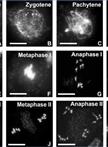
This protocol is a more detailed version of previous protocols (Yang et al., 2011; Bolaños-Villegas et al., 2013) developed for the examination of meiotic chromosome spreads. Meiotic chromosome spreads are useful to determine the presence of defects in chromosome pairing and segregation. The protocol also describes how to perform fluorescent in situ hybridization experiments with a centromere probe used to label chromosomes.