
- Submit a Protocol
- Receive Our Alerts
- EN

- Protocols

- Articles and Issues
- For Authors
- About
- Become a Reviewer

Transmission Electron Microscopy for Tobacco Chloroplast Ultrastructure

Published: Vol 5, Iss 4, Feb 20, 2015 DOI: 10.21769/BioProtoc.1404 Views: 10032
Reviewed by: Tie LiuAnonymous reviewer(s)
Original research article
The authors used this protocol in:
May 2014

Protocol Collections
Comprehensive collections of detailed, peer-reviewed protocols focusing on specific topics


Related protocols
Abstract
The chloroplast is the site of photosynthesis that enabled and sustains aerobic life on Earth. Chloroplasts are relatively large organelles with a diameter of ~5 μm and width of ~2.5 μm, and so can be readily analysed by electron microscopy. Each chloroplast is enclosed by two envelope membranes, which encompass an aqueous matrix, the stroma and the thylakoids. Components of stroma include starch granules and plastoglobuli, which can be observed by electron microscopy. And the thylakoids consist of stromal thylakoid, granal thylakoid and as well as granum (a stack of thylakoids). These structure components are quite sensitive to developmental changes and environmental variations, such as drought, salinity, cold, high temperature and others. Transmission electron microscopy (TEM) is a powerful technique for monitoring the effects of various changing parameters or treatments on the development and differentiation of these important organelles. Here we describe a reliable method for the analysis of plastid ultrastructure in tobacco plant by TEM.
Keywords: ChloroplastBackground
Materials and Reagents
- Tobacco (Nicotiana tabacum) plants (about 6-week grew on MS agar plates, 3-week grew in 1/4 Hoagland solution, and 2~3 weeks grew on soil)
- Glutaraldehyde (Wako Pure Chemical Industries, catalog number: 071-01931 )
- Osmium tetroxide (Nisshin EM Corporation, catalog number: 300 )
- Ethanol (Wako Pure Chemical Industries, catalog number: 057-00456 )
- Propylene oxide (Wako Pure Chemical Industries, catalog number: 165-05026 )
- Quetol 812 set (Nisshin EM Corporation, catalog number: 340 )
- Uranyl acetate (Wako Pure Chemical Industries, catalog number: 6159-44-0 )
- 3% (w/v) lead citrate (Wako Pure Chemical Industries, catalog number: 121-01722 )
- Dodecenyl succinic anhydride (DDSA)
- Methyl nadic anhydride (MNA)
- DMP
- 0.1 M phosphate buffered saline (see Recipes)
- 1% osmium tetroxide (see Recipes)
- Quetol-821 resin (see Recipes)
- Hoagland solution (see Recipes)
Equipment
- Razor blade
- Lens tissue
- Scissors
- Tweezers
- Needle and thread
- Glass tube
- Vacuum equipment
- Balance
- Petri dish
- Oven at 60 °C
- Plastic flat embedding mold (catalog number: 70900 )
- Beaker
- Ultramicrotome (RMC, model: MT-7000 )
- Copper grid
- Transmission electron microscope (JEOL, model: JEM-100CX II )
Procedure
- Cut and trim tobacco leaf with a razor blade to 2 mm x 4 mm size, cover the leaf with lens tissue and tie it with needle and spread which can protect the sample and facilitate the fixation and the following steps (because the sample is easily floating and covered with lens tissue will facilitate sinking during fixation), then put it into 2% glutaraldehyde in a glass tube.
- Vacuum the glutaraldehyde tube by vacuum equipment until no air bubble is coming out, and exhaust slowly to let the fix solution go into leaf tissue.
- Fix in 2% glutaraldehyde for 2 h at room temperature.
- Wash with 0.1 M PBS for 4~5 times and each time for 20~30 min.
- Fix in 1% osmium tetroxide for 2 h.
- Wash with distilled water for 30 min during which change the distilled water 5~8 times.
- Add 50% ethanol and incubate for 10 min.
- Discard 50% ethanol, add 70% ethanol and seal with Parafilm, keep overnight.
- Discard 70% ethanol, add 90% ethanol for 10 min.
- Discard 90% ethanol, wash with 100% ethanol for 3 times and each time for 10 min.
- Add propylene oxide for 2 times and each time for 5 min.
- Put the leaf sample into propylene oxide: Quetol 812 resin = 1:1 (v/v) and covered with aluminum foil overnight.
- Change to Quetol 812 resin and keep for 24 h.
- Embed the leaf sample horizontally with Quetol 812 resin on the Plastic flat embedding mold, and put into 60 °C for 48 h.
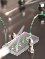
- Cut to thin sections (70~100 nm) with a diamond knife on an ultramicrotome, and the thin sections are gathered on a copper grid which is specially used for transmission electron microscope observation (Figure 1).

Figure 1. A picture of copper grid with sample sections. The red circle indicated the sample sections.
- Stain with 2% (w/v) uranyl acetate for 40 min by putting copper grid with the sample (the sample side is downward) in staining solution and keep for 40 min, the following steps 16-18 are same.
- Wash with distilled water in a beaker for 3 times and each time for 10 min gently.
- Stain with 3% (w/v) lead citrate for 2~3 min.
- Wash with distilled water in a beaker for 3 times and each time for 10 min gently.
- Put on filter paper to make it dry and keep into the case.
- Observe the samples on a transmission electron microscope at 80 kv, and take photos. The sample image of chloroplast can be found in Figure 6 in the Wang et al. (2014) .
Notes
- In step 2, be sure to exhaust slowly to let the fix solution go into the sample tissue.
- During ethanol stepcoppers, keep a small amount of former solution before add new solution, this can avoid the dry of tissue edge.
- When prepare the resin, be sure to mix well before adding DMP-30 accelerator. And the ratio of each reagent is important, but the total amount can be adjusted.
- In steps 15 and 17, stain with uranyl acetate, drop a few drops on a Parafilm in a petridish, and put the copper disc on the drop.
Recipes
- 0.2 M phosphate buffered saline (pH 7.0)
Solution A: 0.2 M Na2HPO4, Na2HPO4.12H2O 7.164 g in 100 ml distilled water
Solution B: 0.2 M NaH2PO4, NaH2PO4.2H2O 3.121 g in 100 ml distilled water
Put 61 ml solution A and 39 ml solution B together and mix well to make 0.2 M phosphate buffered saline at pH 7.0
- 1% osmium tetroxide
4% osmium tetroxide: Distilled water: 0.2 M PBS= 1:1:2
- Quetol-821 resin (in the case of prepare 102 g)
Quetol-812
48 g
DDSA
19 g
MNA
33 g
DMP
302 g
- Hoagland solution
Note: Recipe can be found in Epstein (1972).
Acknowledgments
This work was supported by the National Natural Science Foundation of China (grant no. 31200206), the West Light Foundation of the Chinese Academy of Sciences, and the Chinese Universities Scientific Fund (grant no. ZD2012023).
References
- Epstein, E. (1972). Mineral nutrition of plants: principles and perspectives.
- Wang, S., Uddin, M. I., Tanaka, K., Yin, L., Shi, Z., Qi, Y., Mano, J., Matsui, K., Shimomura, N., Sakaki, T., Deng, X. and Zhang, S. (2014). Maintenance of chloroplast structure and function by overexpression of the rice MONOGALACTOSYLDIACYLGLYCEROL SYNTHASE gene leads to enhanced salt tolerance in tobacco. Plant Physiol 165(3): 1144-1155.
Article Information
Copyright
© 2015 The Authors; exclusive licensee Bio-protocol LLC.
How to cite
Readers should cite both the Bio-protocol article and the original research article where this protocol was used:
- Yin, L., Wang, S., Shimomura, N. and Tanaka, K. (2015). Transmission Electron Microscopy for Tobacco Chloroplast Ultrastructure. Bio-protocol 5(4): e1404. DOI: 10.21769/BioProtoc.1404.
-
Wang, S., Uddin, M. I., Tanaka, K., Yin, L., Shi, Z., Qi, Y., Mano, J., Matsui, K., Shimomura, N., Sakaki, T., Deng, X. and Zhang, S. (2014). Maintenance of chloroplast structure and function by overexpression of the rice MONOGALACTOSYLDIACYLGLYCEROL SYNTHASE gene leads to enhanced salt tolerance in tobacco. Plant Physiol 165(3): 1144-1155.
Category
Plant Science > Plant physiology > Photosynthesis
Plant Science > Plant cell biology > Cell structure
Cell Biology > Cell imaging > Electron microscopy
Do you have any questions about this protocol?
Post your question to gather feedback from the community. We will also invite the authors of this article to respond.
![]() Tips for asking effective questions
Tips for asking effective questions
+ Description
Write a detailed description. Include all information that will help others answer your question including experimental processes, conditions, and relevant images.