
- Submit a Protocol
- Receive Our Alerts
- EN

- Protocols

- Articles and Issues
- For Authors
- About
- Become a Reviewer

Rapid Profiling Cell Cycle by Flow Cytometry Using Concurrent Staining of DNA and Mitotic Markers
Published: Vol 7, Iss 16, Aug 20, 2017 DOI: 10.21769/BioProtoc.2517 Views: 11840
Reviewed by: Emilie BesnardAnonymous reviewer(s)
Original research article
The authors used this protocol in:

Sep 2015

Protocol Collections
Comprehensive collections of detailed, peer-reviewed protocols focusing on specific topics


Abstract
The flow cytometric quantitation of DNA content by DNA-binding fluorochrome, propidium iodide (PI) is the most widely used method for cell cycle analysis. However, the commonly used methods are time-consuming and labor-intensive and are incompatible with staining of mitotic markers by fluorescent-labeled antibodies. Here, we report an optimized simple protocol for rapid and simultaneous analysis of characteristic mitotic phosphorylated proteins and DNA content, permitting the quantification of cells in mitosis, G1, S and G2 stage in a single assay. The protocol detailed here employs detergent-based hypotonic solution to rapidly permeabilize cells and allows simultaneous staining of DNA with PI and mitotic marker, phospho-Histone H3, with specific antibody within 20 min. The protocol requires only inexpensive and commercial available reagents and also enables a rapid and complete analysis of cell cycle profile.
Keywords: Cell cycleBackground
Cell cycle analysis by flow cytometry is mainly based on measurement of DNA content by stain with propidium iodide (PI). The stoichiometric nature of PI ensures the accurate quantification of DNA content and allows us to reveal the distribution of cells in G1, S and G2 cell cycle stage or in sub-G1 cell death stage, the latter of which is characterized by DNA fragmentation. Most of the commonly used methods for PI-based DNA quantification require fixation using alcohol or aldehyde, which is time-consuming and labor-intensive. In addition, these methods are incompatible with staining of mitotic markers by fluorescent-labeled antibodies. Therefore, we adopted and optimized a previous established hypotonic buffer to permeabilize cells, allowing simultaneous staining of DNA with PI and mitotic marker, phospho-Histone H3 (pH3), with pH3 specific antibody (Riccardi et al., 2006; Liu et al., 2016). This method enables a rapid (within 20 min) and comprehensive analysis of cell cycle profile (G1, S, G2 and M phase).
Materials and Reagents
- Falcon® 5 ml round bottom polystyrene test tube (Corning, Falcon®, catalog number: 352058 )
- Dulbecco’s phosphate buffered saline, no calcium, no magnesium (DPBS) (Thermo Fisher Scientific, Thermo ScientificTM, catalog number: 14200166 )
- Alexa Fluor® 647 rat anti-phospho-Histone H3 (pS28) (BD, BD Bioscience, catalog number: 558217 )
- Sodium citrate (Na3C6H5O7·2H2O) (Sigma-Aldrich, catalog number: 1613859 )
- TritonTM X-100 (Sigma-Aldrich, catalog number: T8787 )
- Propidium iodide (PI) (C27H34I2N4) (Sigma-Aldrich, catalog number: P4170 )
- Hypotonic lysis/PI buffer (see Recipes)
Equipment
- Centrifuge (Eppendorf, model: 5424 R )
- Flow cytometer (ACEA BIO, model: NovoCyte Flow Cytometer , 488-nm laser line, for excitation)
Software
- FlowJo_V10 is used for analyzing the flow cytometry data
Procedure
Note: All the procedures are done at room temperature.
- Suspend cells at 0.4 x 10⁶ cells in 1 ml of DPBS in Falcon® 5 ml tubes.
- Centrifuge at 200 x g for 5 min.
- Aspirate off the DPBS.
- Re-suspend the cell pellet in 100 μl of hypotonic buffer, add 0.5 μl Alexa Fluor® 647 anti-phospho-Histone H3 per sample (Alexa Fluor 647 can be detected at APC channel), then gently mix the sample.
Note: Since the hypotonic shock removes the majority of the RNA, RNase treatment is not required. - Place the tubes in the dark at room temperature, before flow cytometry analysis, for at least 20 min but no longer than 2 h.
Note: Including an additional washing step before measurement does not change the result but may cause cell loss. - Measure fluorescence (detector 585/40 nm at PE channel for PI and 675/30 nm at APC channel for phospho-Histone H3 staining) by flow cytometer. Collect at least 20,000 total events, gate-out residual debris and measure diploid and tetraploid DNA peaks (Figures 1A-1D).
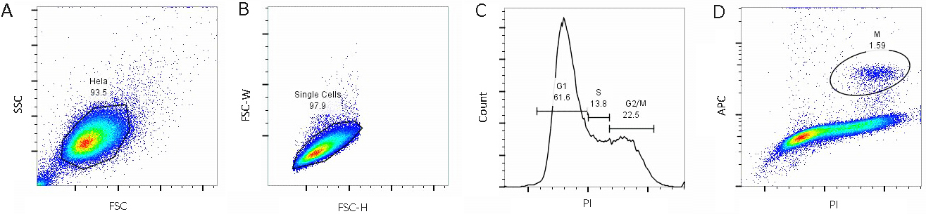
Figure 1. Illustration of the gating strategy in analyzing cell cycle profile of HeLa cell. A. Exclusion of cell debris by forward scatter (FSC) and side scatter (SSC); B. Exclusion of clumps and doublets by forward scatter width (FSC-W) and height (FSC-H); C. Identification of G1, S and G2/M phase by PI; D. Identification of M-phase by mitosis-specific antibody (phospho-Histone H3-APC). The final percentage (%) of cells for each phase is about G1: 61.6%, S: 13.8%, G2/M: 22.5, M: 1.59% according to the analysis.
Data analysis
As shown in Figure 1, the histogram of DNA content could categorize cells into three groups, G1, S and G2/M phase (Figure 1C). Mitosis-specific antibody (phospho-Histone H3-APC) is applied to distinguish G2 from M cells (Figure 1D).
Notes
- For applying this protocol to other cell lines, the careful titration on cell number is recommended.
- Leaving the cell in hypotonic buffer for longer than 2 h may significantly increase the cell debris.
- Hypotonic buffer is not compatible with concurrent examination of intracellular fluorescent (GFP, RFP etc.) fusion proteins.
Recipes
- Hypotonic lysis/PI buffer
Deionized/distilled water
0.1% sodium citrate (wt/v)
0.1% Triton X-100 (v/v)
50 μg/ml PI
Notes:- Solution can be kept at 4 °C for months.
- PI is a suspected carcinogen and should be handled with care. The dye must be disposed of safely and in accordance with applicable local regulations.
- Solution can be kept at 4 °C for months.
Acknowledgments
This work was supported by R21AI117547 and 1R01AI114581 from National Institute of Health, V2014-001 from the V-Foundation and 128436-RSG-15-180-01-LIB from the American Cancer Society (R.W.).
References
- Liu, L., Lu, Y., Martinez, J., Bi, Y., Lian, G., Wang, T., Milasta, S., Wang, J., Yang, M., Liu, G., Green, D. R. and Wang, R. (2016). Proinflammatory signal suppresses proliferation and shifts macrophage metabolism from Myc-dependent to HIF1α-dependent. Proc Natl Acad Sci U S A 113(6): 1564-1569.
- Riccardi, C. and Nicoletti, I. (2006). Analysis of apoptosis by propidium iodide staining and flow cytometry. Nat Protoc 1(3): 1458-1461.
Article Information
Copyright
© 2017 The Authors; exclusive licensee Bio-protocol LLC.
How to cite
Shen, Y., Vignali, P. and Wang, R. (2017). Rapid Profiling Cell Cycle by Flow Cytometry Using Concurrent Staining of DNA and Mitotic Markers. Bio-protocol 7(16): e2517. DOI: 10.21769/BioProtoc.2517.
Category
Cancer Biology > Cell cycle checkpoints > Cell-based assays
Cell Biology > Cell-based analysis > Flow cytometry
Do you have any questions about this protocol?
Post your question to gather feedback from the community. We will also invite the authors of this article to respond.
![]() Tips for asking effective questions
Tips for asking effective questions
+ Description
Write a detailed description. Include all information that will help others answer your question including experimental processes, conditions, and relevant images.